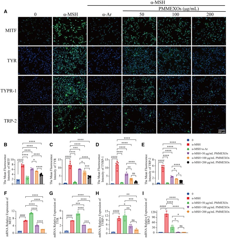
IMAGE

Figure 4.

ID
ZDB-IMAGE-250801-96
Source
Figures for Mo et al., 2025
Image
Figure Caption

Figure 4.

PMMEXOs inhibits the expression of melanin-related proteins and genes. (A) Immunofluorescence staining of MITF, TYR, TYRP-1 and TRP-2 proteins during melanin production. After incubation for 24 h, fluorescent of MITF, TYR, TYRP-1 and TRP-2 proteins, nuclei stained with DAPI. Scale bars = 200 μm. Quantitative fluorescence intensity analysis of MITF (B), TYR (C), TYRP-1 (D), TRP-2 (E) proteins. n = 5. The mRNA expression levels of MITF (F), TYR (G), TYRP-1 (H) and TRP-2 (I) in PMMEXOs treated cells were detected by RT-qPCR (n = 3). Data were expressed as mean ± SD, and one-way analysis of variance was used for statistical significance. n. s. no significant difference observed between groups. *P < 0.05, **P < 0.01, ***P < 0.001, ****P < 0.0001 compared with α-MSH model group, ####P < 0.0001 compared with the blank group.

Acknowledgments
This image is the copyrighted work of the attributed author or publisher, and ZFIN has permission only to display this image to its users. Additional permissions should be obtained from the applicable author or publisher of the image. Full text @ Regen Biomater