IMAGE

Figure 2.

ID
ZDB-IMAGE-250801-93
Source
Figures for Mo et al., 2025
Image
Figure Caption

Figure 2.

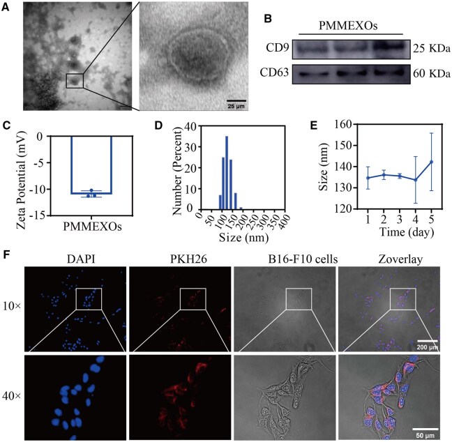
Characterization of PMMEXOs. (A) Morphology of PMMEXOs, observed by transmission electron microscopy (TEM). Scale bars = 25 μm. (B) Western blot analysis of EV biomarkers in PMMEXOs, including CD63 and CD9. (C) DLS represents the particle zeta of PMMEXOs. (D) DLS indicates the particle size of PMMEXOs. (E) Storage stability: PMMEXOs was stored at pH7.4 PBS, −80°C for 5 days and the particle size was determined. (F) The absorption of PMMEXOs by B16-F10 cells was observed by micrograph (magnification: 10 ×, scale bars = 200 μm) and magnification (magnification: 40 ×, scale bars = 50 μm). After incubation for 24 h, fluorescent PKH26-labeled PMMEXOs, nuclei stained with DAPI. n = 3.

Acknowledgments
This image is the copyrighted work of the attributed author or publisher, and ZFIN has permission only to display this image to its users. Additional permissions should be obtained from the applicable author or publisher of the image. Full text @ Regen Biomater