IMAGE

Figure 9.

ID
ZDB-IMAGE-250801-102
Source
Figures for Mo et al., 2025
Image
Figure Caption

Figure 9.

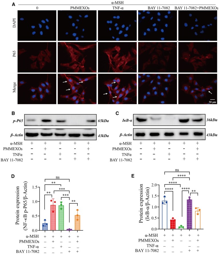
PMMEXOs reduces melanin production by activating the NF-κB signaling pathway. (A) The translocation of NF-κB p-65 from cytoplasm to nucleus of B16-F10 cells after different drug stimulation was detected by immunofluorescence assay. The arrow indicates the translocation of NF-κB p-65 from cytoplasm to nucleus. Fluorescent of NF-κB p-65 proteins, nuclei stained with DAPI. Scale bars = 50 μm. WB detection of NF-κB p-P65 (B) and IκB-α (C) protein expression. The relative protein expression level of NF-κB p-P65 (D) and IκB-α (E) were quantitatively analyzed. n = 3. Data are expressed as mean ± SD, one-way analysis of variance was used for statistical significance. n. s. no significant difference observed between groups. *P < 0.05, **P < 0. 01, ***P < 0.001, ****P < 0.0001.

Acknowledgments
This image is the copyrighted work of the attributed author or publisher, and ZFIN has permission only to display this image to its users. Additional permissions should be obtained from the applicable author or publisher of the image. Full text @ Regen Biomater