
IMAGE
Fig. 4
- ID
- ZDB-IMAGE-250728-77
- Publication
- Gregory-Evans et al., 2025 - Mutation of beta-tubulin 4B gene (TUBB4B) causes autosomal dominant retinitis pigmentosa with sensorineural hearing loss in a multigenerational family
- All Figures
- Figures for Gregory-Evans et al., 2025
Image
|
Figure Caption
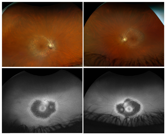
Fig. 4 Color fundus and autofluorescence images from affected participant III-1. The upper row shows color fundus images from age 63 years showing bone spicule retinal degeneration confined to the near periphery. The bottom row shows autofluorescence imaging showing a hypo-autofluorescence band confined to the posterior pole plus hyper-autofluorescence beyond this area and at each macula. This would be described as pericentral retinitis pigmentosa.
Acknowledgments
This image is the copyrighted work of the attributed author or publisher, and
ZFIN has permission only to display this image to its users.
Additional permissions should be obtained from the applicable author or publisher of the image.