
|
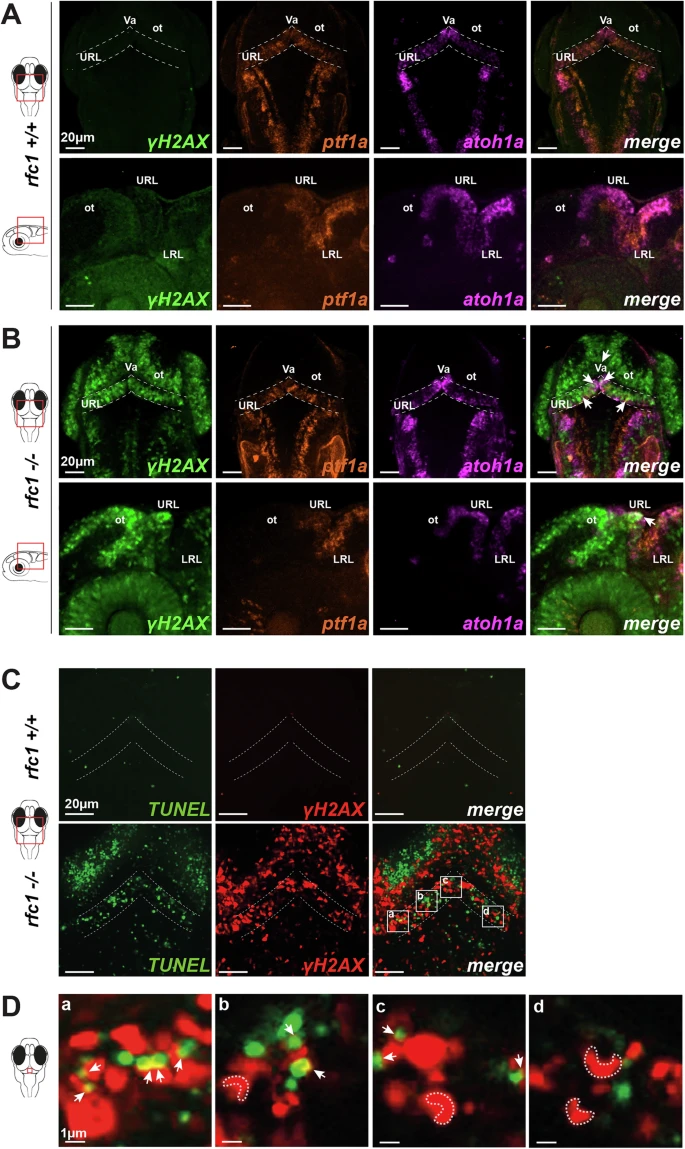
Fig. 6 Cerebellar neural progenitors accumulate DNA damage and undergo apoptosis in the absence of RFC1.A, B Combined immunofluorescence and RNAscope in situ hybridization for DNA damage marker γH2AX (green), ptf1a (orange), and atoh1a (magenta) at 2 dpf in rfc1+/+ (A, n = 5) and rfc1−/− (B, n = 5) embryos. Dorsal views (upper panels) and lateral views (lower panels) are shown. Dotted lines outline the upper rhombic lip (URL). ot optic tectum, LRL lower rhombic lip, Va valvula cerebelli. C Confocal images (1-μm optical slice) showing colocalization of apoptotic cells (TUNEL, green) and DNA damage (γH2AX, red) at 2 dpf in rfc1+/+ (n = 5) and rfc1-/- (n = 5) embryos. Dotted lines delineate the cerebellar region. D Higher magnification of selected regions from (C) (a–d), highlighting colocalization of TUNEL and γH2AX signals (white arrows) and characteristic γH2AX apoptotic ring patterns (dotted outlines).