IMAGE

Fig. 7

ID
ZDB-IMAGE-250724-44
Source
Figures for Herdt et al., 2025
Image
Figure Caption

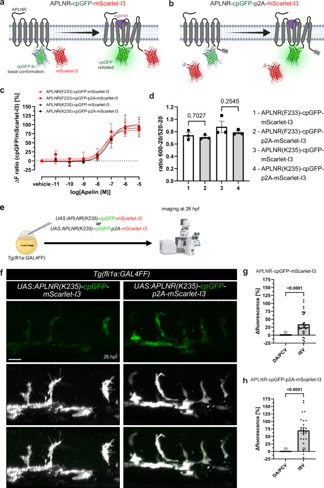
Fig. 7 Development and in vivo application of ratiometric APLNR-cpGFP-mScarlet-I3 biosensors.

a, b Schematic illustration of the ratiometric APLNR conformational biosensors design. mScarlet-I3 and p2A-mScarlet-I3 were integrated C-terminally into the APLNR-cpGFP biosensors, resulting in membrane-bound (a) and cytosolic (b) mScarlet-I3 localization. c HEK293A cells were transiently transfected with the respective ratiometric biosensor plasmid and stimulated with Apelin. Stimulation with Apelin led to a strong increase in cpGFP over mScarlet-I3 fluorescence intensity of each ratiometric biosensor variant. d Quantification of the FRET ratio of the ratiometric biosensor with and without a p2A self-cleaving peptide exhibited no significant differences, indicating that cpGFP and mScarlet-I3 are no FRET partner. e Schematic illustration of the procedure. Plasmids encoding either UAS:APLNR(K235)-cpGFP-mScarlet-I3 or UAS:APLNR(K235)-cpGFP-p2A-mScarlet-I3 were injected into 1-cell stage Tg(fli1a:GAL4FF) zebrafish embryos and imaged at 28 hours post fertilization (hpf). f, g Quantification of APLNR(K235)-cpGFP over mScarlet-I3 fluorescence intensities in ISVs compared to the DA and PCV in UAS:APLNR(K235)-cpGFP-mScarlet-I3 (g) or UAS:APLNR(K235)-cpGFP-p2A-mScarlet-I3 (h) injected embryos at 28 hpf. Data in are presented as mean values ± SEM from three independent experiments conducted in transiently transfected HEK293A cells (c, d) or zebrafish embryos (N number of embryos, n either ISVs or DA/PCV: g DA/PCV N/n: 32/32, ISV N/n: 32/110; h DA/PCV N/n: 25/25, ISV N/n: 25/75). Statistical analysis was performed by using ordinary One-way ANOVA, followed Dunnett’s multiple comparison correction (d) and two-tailed unpaired Student’s t-test with Welch’s correction (g, h). Scale bars 50 µm. ISV intersegmental vessel, DA dorsal aorta, PCV posterior cardinal vein, hpf hours post-fertilization, cpGFP circularly permuted GFP, HEK293 human embryonic kidney 293. Source data are provided as a Source Data file. a, b Created in BioRender. Schihada (2025) https://BioRender.com/odhtcw2. e Created in BioRender. Schihada (2025) https://BioRender.com/mz23zor.

Acknowledgments
This image is the copyrighted work of the attributed author or publisher, and ZFIN has permission only to display this image to its users. Additional permissions should be obtained from the applicable author or publisher of the image. Full text @ Nat. Commun.