
Fig. 3
- ID
- ZDB-IMAGE-250724-40
- Publication
- Herdt et al., 2025 - In vivo measurement of an Apelin gradient with a genetically encoded APLNR conformation biosensor
- All Figures
- Figures for Herdt et al., 2025
|
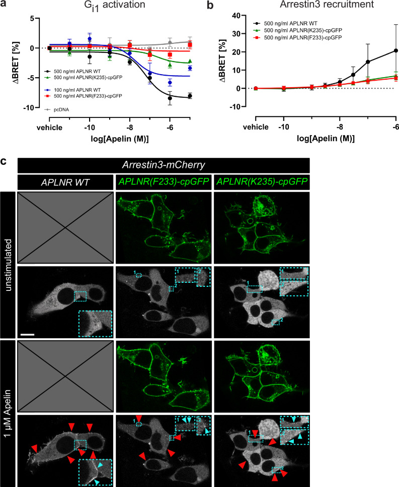
Fig. 3 APLNR-cpGFP biosensors possess signaling ability.
Gi1 protein dissociation (