Image
Figure Caption
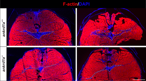
Fig. 9 Representative examples of wounded skeletal muscle tissue in wild-type (wt) and ankrd1a mutant zebrafish at 7 days postinjury (dpi). Injury is recognized as an area without phalloidin-stained myofibers (red) and with nuclei accumulation (blue). Imaging was performed on sections of ankrd1a−/− and ankrd1a+/+ zebrafish (n = 4), which were age-matched and injured at the same time. White dashed lines delineate the remaining injured muscle tissue. Scale bar, 400 µm.
Acknowledgments
This image is the copyrighted work of the attributed author or publisher, and
ZFIN has permission only to display this image to its users.
Additional permissions should be obtained from the applicable author or publisher of the image.
Full text @ Am. J. Physiol. Cell Physiol.

