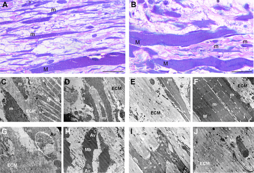
Fig. 8 Histology and ultrastructure of the injury area in ankrd1a mutant and wild-type (wt) skeletal muscle at 5 days postinjury (dpi). Representative light microscopy (LM) images of ankrd1a+/+ (A) and ankrd1a−/− (B) skeletal muscle (n = 4). Muscle fibers are blue, collagen is pink. C–F: representative transmission electron microscopy (TEM) images of ankrd1a+/+ (C and D) and ankrd1a−/− (E and F) skeletal muscle. G–I: autophagic vesicles observed in ankrd1a+/+ (G) and ankrd1a−/− (H and I) skeletal muscle. J: macrophage detected in ankrd1a−/− skeletal muscle. Av and *, autophagy vesicle; ECM, extracellular matrix; M, myofiber; m, newly growing myofiber; Mb, myoblast; Mφ, macrophage. Scale bars, 20 µm for A and B and 1 µm for C and J.
Image
Figure Caption
Acknowledgments
This image is the copyrighted work of the attributed author or publisher, and
ZFIN has permission only to display this image to its users.
Additional permissions should be obtained from the applicable author or publisher of the image.
Full text @ Am. J. Physiol. Cell Physiol.

