
|
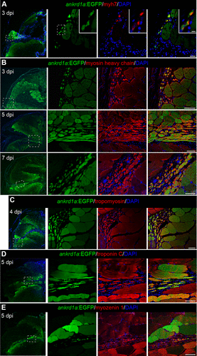
Fig. 6 Newly forming myofibers express TgBAC(ankrd1a:EGFP). Cryosections of injured skeletal muscle of the reporter line TgBAC(ankrd1a:EGFP) (n = 3), at indicated timepoints, were stained for muscle markers: embryonic myosin Myh7 (A), myosin (B), tropomyosin (C), troponin C (D), and myozenin 1 (E). Nuclei were stained with DAPI. Low-magnification images are on the left with marked positions (dashed boxes) of higher-magnification images on the right. Elongated myofibers inside the injury area on B and D correspond to longitudinally cut fibers. Scale bars, 20 μm for A, 5 μm for insets in A, and 50 μm for B–E, for higher magnification images on the right. EGFP, enhanced green fluorescent protein.