
|
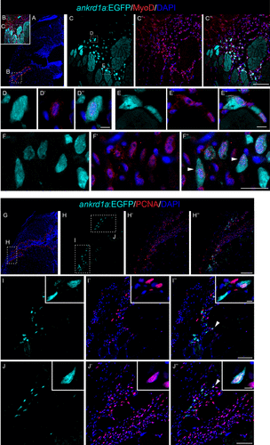
Fig. 5 Myoblast-like (A–F″) and proliferating (G–J″) cells express TgBAC(ankrd1a:EGFP) in injured zebrafish skeletal muscle. Cryosections of zebrafish muscle at 4 days postinjury (dpi) (n = 3) were subjected to immunostaining for Myod1 and Pcna. A and G: low magnification images of the representative muscle section stained with DAPI, with delineated injury region (red dashed line) and positions of higher magnification images (C–C″ and H–H″). White arrowheads on F″—EGFP+ myoblast-like cells. White arrowheads on I″ and J″ point to cells magnified in insets. Scale bars, 100 μm for H–H″; 50 μm for C–C″, I–I″, and J–J″; 25 μm for F–F″; 5 μm for D–D″, E–E″, and insets in I and J. EGFP, enhanced green fluorescent protein.