
|
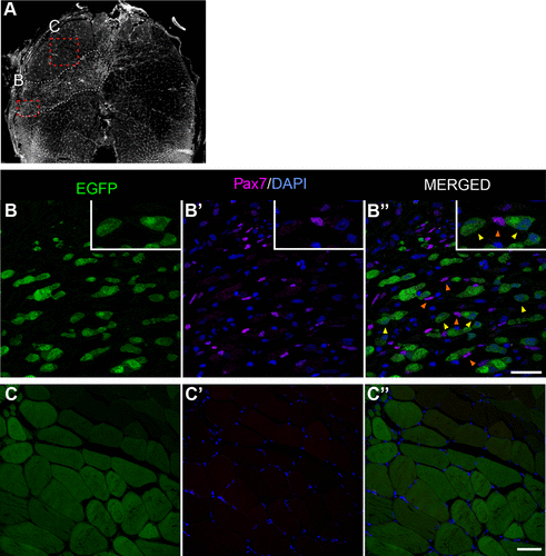
Fig. 4 TgBAC(ankrd1a:EGFP) expression was not detected in activated satellite-like cells found within the injury area. Cryosections of injured skeletal muscle at 4 days postinjury (dpi) (n = 3) were immunostained with anti-Pax7 antibody, and nuclei were labeled with DAPI. A: low magnification image of the representative muscle section stained with DAPI, with delineated injury region (white dashed line) and positions of higher magnification images in injured (B–B″) and uninjured (C–C″) regions. Orange arrowheads on B″ point to some of the Pax7-positive nuclei as examples. Yellow arrowheads point to Pax7-negative myoblasts with visible nuclei. No Pax7-positive nuclei were detected in the adjacent uninjured region. Scale bars, 25 μm for B–B″ and 50 μm for C–C″. EGFP, enhanced green fluorescent protein.