
|
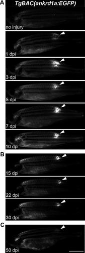
Fig. 2 Expression of TgBAC(ankrd1a:EGFP) in injured skeletal muscle of adult zebrafish. А: three zebrafish per each timepoint were injured and left to recover for 1, 3, 5, 7, and 10 days before imaging. B: for longer timepoints, 3 zebrafish were injured. At 15 and 22 days postinjury (dpi), they were anaesthetized, imaged, and returned to the system water. At 30 dpi, they were euthanized, images were acquired, and tissue around the injury was used for RNA isolation. C: three zebrafish were injured, left to recover for 50 days, and imaged. Images of one representative zebrafish per experiment are shown. White arrowheads point to the fluorescence signal at the site of injury. The images on the same panel were acquired under the same conditions. Scale bar, 5 mm. EGFP, enhanced green fluorescent protein.