
|
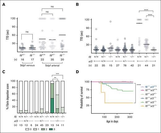
Fig. 2 f8 mutants show an altered hemostatic balance compared to mammals. (A) FVIII-deficient zebrafish laser-mediated endothelial injury of the PCV was performed on larvae at 3 dpf, and dorsal aorta at 5 dpf. The TTO was not significantly different in f8–/– compared to f8+/− clutchmates (Mann-Whitney U test P > 1). Circles represent individual larvae. Horizontal bars represent the median TTO. (B) Laser-mediated injury on f8+/−;at3+/− incrosses reveal that loss of FVIII reverses the at3–/– phenotype. In the at3–/– background, the increased TTO is reversed by mutation of f8 in a dose-dependent fashion (∗∗∗∗P < .001 by Mann-Whitney U testing). (C) Fibrin deposition observed in 5 dpf fgb-egfp larvae resulting from f8+/−;at3+/− incrosses. Bar graph represents the percentage of larvae in each fibrin deposition category: score 0 having no GFP-labeled fibrin deposits in the PCV, score 1 with <5 occurrences, score 2 with 5 to 25 occurrences, and score of 3 with widespread continuous threads of fibrin in the PCV and/or surrounding regions. Overall statistical significance was determined by Kruskal-Wallis and pairwise comparisons among the f8+/+;at3–/–, f8+/−;at3–/–, and f8–/–;at3–/– mean fibrin scores by a Wilcoxon rank sum test with a Bonferroni correction (∗∗P < .005; ∗∗∗P < .0005). (D) Survival curve of zebrafish offspring from f8+/−;at3+/− incrosses shows that loss of f8 rescues the at3–/– lethal phenotype with a statistically significant difference between f8+/+;at3–/– and f8–/–;at3–/– (∗∗∗∗P < .0001 by log-rank Mantel-Cox testing). f8+/+;at3+/+ n = 22, f8+/−;at3+/− n = 83, f8+/+;at3–/– n = 3, f8+/−;at3–/–, n = 27, f8–/–;at3–/–, n = 22, f8–/–;at3+/+ n = 18. ns, nonsignificant.