
|
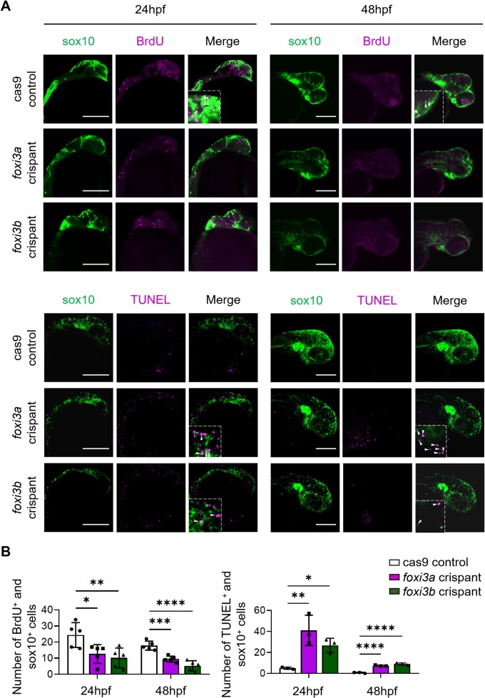
Fig. 4 Cranial neural crest cells in foxi3a and foxi3b crispants display decreased proliferation and increased apoptosis. A. Confocal images of proliferating cells labeled with BrdU staining (magenta) and of apoptotic cells labeled with TUNEL staining (magenta) showing sox10 positive (green) CNCC proliferation decreases and apoptosis increases in foxi3a and foxi3b Tg(sox10:GFP) crispants at 24 hours postfertilization (hpf). Scale bars, 200 μm. B. Quantification of BrdU- and TUNEL-positive CNCCs in cas9 controls and foxi3a and foxi3b crispants at 24 hpf (n ≥ 3 larvae per group for each time point; *p < 0.05, **p < 0.01, ***p < 0.001, and ****p < 0.0001; one way ANOVA, Dunnett test). All error bars indicate mean ± standard deviation.