IMAGE

Fig 4

ID
ZDB-IMAGE-250711-19
Source
Figures for Ravishankar et al., 2025
Image
Figure Caption

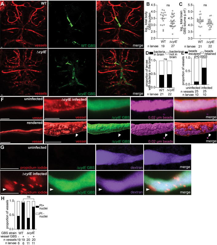
Fig 4 Endothelial cell lysis occurs independently of the primary GBS cytolysin, cylE.

(A) Representative confocal images of red fluorescent brain vasculature in 20 hpi larvae infected with approximately 50 CFU wildtype (WT) GBS-GFP (top), or approximately 150 CFU ∆cylE GBS-GFP (bottom). (B) Wildtype or ∆cylE burden per larva at 20 hpi quantified by FPC. Horizontal bars, means; ns: not significant, Student t test. Representative of 3 independent experiments. (C) Quantification of total GBS volume in the brain of wildtype or ∆cylE GBS infected larvae. Horizontal bars, means; ns: not significant, Student t test. Representative of 3 independent experiments. (D) Proportion of wildtype or ∆cylE GBS-infected larvae with GBS in the brain; ns: not significant, Fisher’s exact test. Representative of 3 independent experiments. (E) Proportion of uninfected or ∆cylE GBS-infected vessels with beads escaped or retained in the vessels; Fisher’s exact test. (F) Representative confocal image (top) and 3D rendering (bottom) of an uninfected (left) and infected (right) brain blood vessel from a 20 hpi larva infected with approximately 150 CFU ∆cylE GBS-GFP and injected intravenously with 0.02 µm far red beads (pseudocolored magenta). White arrowheads, escaped beads. (G) Representative confocal images of an uninfected (top) and infected (bottom) purple fluorescent (from injected dextran, pseudocolored magenta) brain blood vessel. Larvae were infected with approximately 150 CFU ∆cylE GBS-GFP and injected intravenously with red fluorescent propidium iodide (PI) just prior to imaging at 20 hpi. White arrowhead, PI positive endothelial cell nucleus. (H) Proportion of uninfected and wildtype or ∆cylE GBS infected vessels with PI-positive nuclei; ns: not significant, Fisher’s exact test. Scale bar, 10 µm throughout. All underlying data in Fig 4 can be found in the supplemental Excel file entitled “S1 Data”.

Acknowledgments
This image is the copyrighted work of the attributed author or publisher, and ZFIN has permission only to display this image to its users. Additional permissions should be obtained from the applicable author or publisher of the image. Full text @ PLoS Biol.