IMAGE

Fig 1

ID
ZDB-IMAGE-250711-15
Source
Figures for Ravishankar et al., 2025
Image
Figure Caption

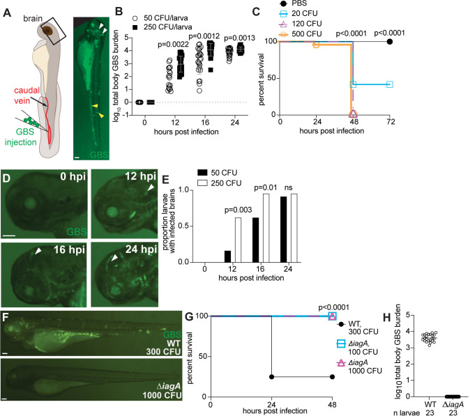
Fig 1 GBS infects the brain of zebrafish larvae in a time- and inoculum-dependent manner.

(A) Diagram of zebrafish caudal vein injection (left). Green fluorescent GBS-GFP infected larva at 20 hours post infection (hpi) (right). White arrowheads, GBS infection in the brain. Yellow arrowheads, GBS infection in the body. (B) GBS burden per larva infected with 50 or 250 CFU at 0–24 hpi, quantified by fluorescent pixel count (FPC). n = 24 larvae per group. Horizontal bars, means; Student t test. Representative of 2 independent experiments. (C) 72-h survival curve of zebrafish larvae injected with 20–500 CFU GBS. n = 24 larvae per group; Kaplan–Meier test, compared to PBS-injected group. Representative of 3 independent experiments. (D) Representative images of the same larva head, infected with 220 CFU GBS-GFP, at 0, 12, 16, and 24 hpi. White arrowheads, GBS infection in brain. (E) Proportion of larvae with GBS-infected brains with 50 CFU (black) or 250 CFU (white) inoculum at 0, 12, 16, and 24 hpi. n = 24 larvae per group; ns: not significant, Fisher’s exact test. Representative of 2 independent experiments. (F) Representative images of larvae infected with 300 CFU wildtype (WT) GBS-GFP, or 1,000 CFU ∆iagA GBS-GFP. (G) 48-hour survival curve of larvae infected with wildtype GBS-GFP or 1,000 CFU ∆iagA GBS-GFP. n = 24 larvae per group; Kaplan–Meier test, compared to wildtype group. Representative of 3 independent experiments. (H) GBS burden per larva infected with 100 CFU wildtype or 1,000 CFU ∆igaA at 24 hpi, quantified by FPC. Horizontal bars, means; Student t test. Scale bar, 100 µm throughout. All underlying data in Fig 1 can be found in the supplemental Excel file entitled “S1 Data”.

Acknowledgments
This image is the copyrighted work of the attributed author or publisher, and ZFIN has permission only to display this image to its users. Additional permissions should be obtained from the applicable author or publisher of the image. Full text @ PLoS Biol.