
Image
|
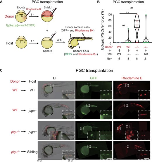
Figure Caption
Fig. 4 Defective PGC migration in pigu mutants is caused by the loss of Pigu in both PGCs and somatic cells. (A) Schematic showing PGC transplantation. (B) The ratios of ectopic PGCs to total transplanted PGCs per embryo in four PGC transplantation pairs. Ne, the number of transplanted embryos. Sib, sibling. –/–, pigu–/–. **P < 0.01. (C) Fluorescence images showing host embryos with successfully transplanted PGCs. BF, bright field. GFP, labeling PGCs. Rhodamine B, labeling donor cells. The dashed boxed areas were enlarged for better review. The arrows indicate ectopically located PGCs.
Acknowledgments
This image is the copyrighted work of the attributed author or publisher, and
ZFIN has permission only to display this image to its users.
Additional permissions should be obtained from the applicable author or publisher of the image.
Full text @ J. Mol. Cell Biol.