IMAGE

Figure 6.

ID
ZDB-IMAGE-250705-24
Source
Figures for Abgueguen et al., 2025
Image
Figure Caption

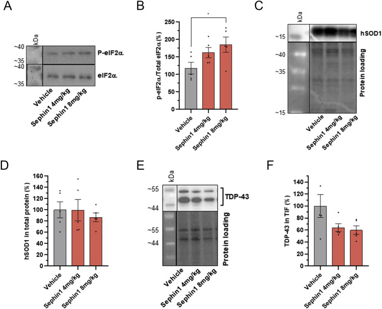
Figure 6. Sephin1 increases eIF2α phosphorylation level and reduces TDP-43 in TIF from spinal cord motor neurons of SOD1G93A female mice at 20 wk of age.

(A) Representative pictures of immunoblot of eIF2α phosphorylated and eIF2α proteins in spinal cord of 20-wk-old SOD1G93A mice treated with vehicle, Sephin1 at 4 or 8 mg/kg per day. (B) eIF2α phosphorylation level in the spinal cord of 20-wk-old SOD1G93A mice treated with vehicle, Sephin1 at 4 or 8 mg/kg per day. (C) Representative images of immunoblot of hSOD1 protein in spinal cord of 20-wk-old SOD1G93A mice treated with vehicle, Sephin1 at 4 or 8 mg/kg per day. (D) hSOD1 protein level in spinal cord of 20-wk-old SOD1G93A mice treated with vehicle, Sephin1 at 4 or 8 mg/kg per day. (E) Representative picture of TDP-43 immunoblot of TIF from spinal cord of 20-wk-old SOD1G93A mice treated with vehicle, Sephin1 at 4 or 8 mg/kg per day. (F) TDP-43 level in TIF from spinal cord of 20-wk-old SOD1G93A mice treated with vehicle, Sephin1 at 4 or 8 mg/kg per day. *P < 0.05 versus vehicle Kruskal-Wallis test followed by Dunn’s multiple comparison test; mean ± SEM n = 3–5 mice per condition.

Source data are available for this figure.

Acknowledgments
This image is the copyrighted work of the attributed author or publisher, and ZFIN has permission only to display this image to its users. Additional permissions should be obtained from the applicable author or publisher of the image. Full text @ Life Sci Alliance