IMAGE

Figure 3.

ID
ZDB-IMAGE-250705-20
Source
Figures for Abgueguen et al., 2025
Image
Figure Caption

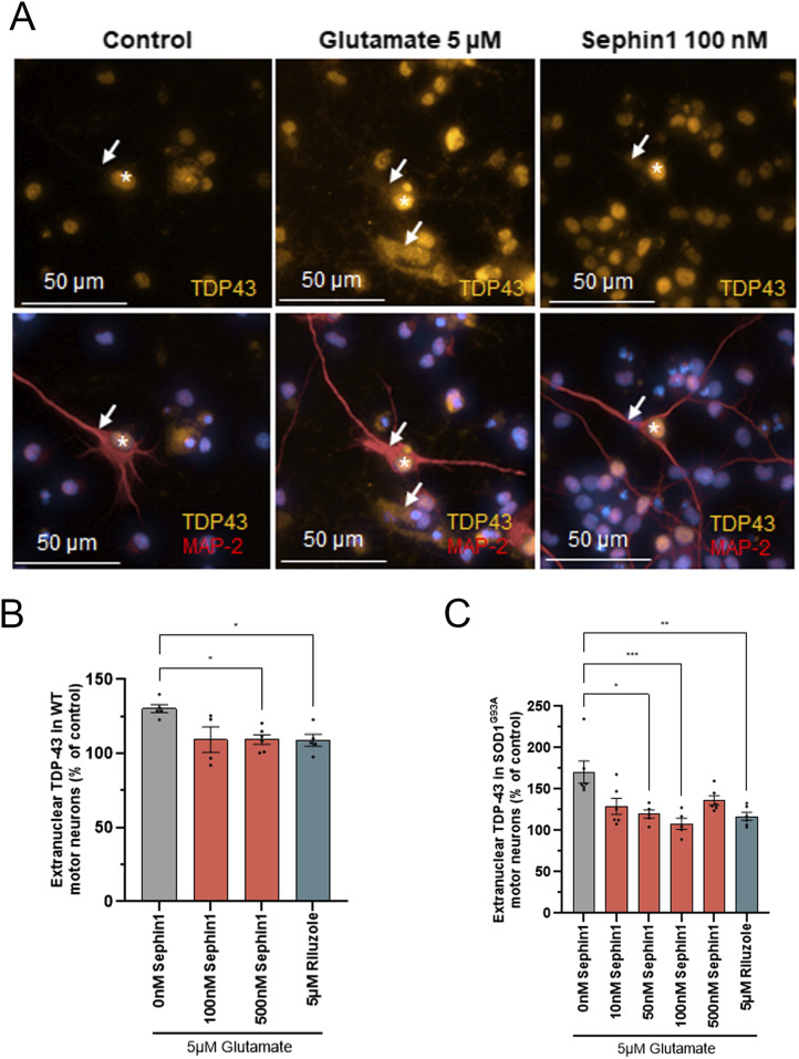
Figure 3. Sephin1 reduces extranuclear TDP-43 localization in primary motor neurons from WT and SOD1G93A rats.

(A) Representative pictures of TDP-43 localization in control primary SOD1G93A rat motor neurons, in primary SOD1G93A rat motor neurons intoxicated by glutamate and in primary SOD1G93A rat motor neurons intoxicated by glutamate in presence of Sephin1 100 nM. White arrows: TDP-43 localization in cytoplasm of MAP-2 labeled motor neurons, white star: TDP-43 localization in nucleus of MAP-2 labeled motor neurons. (B) Extranuclear TDP-43 level in primary WT rat motor neurons intoxicated by glutamate and treated with DMSO, Sephin1, or riluzole 5 μM for 24 h. (C) Extranuclear TDP-43 level in primary SOD1G93A rat motor neurons intoxicated by glutamate and treated with DMSO, Sephin1, or riluzole 5 μM for 24 h *P < 0.05, **P < 0.01, ***P < 0.001 versus 0 nM Sephin1 Kruskal-Wallis test followed by Dunn’s multiple comparison test; mean ± SEM n = 4–6 replicates per condition.

Acknowledgments
This image is the copyrighted work of the attributed author or publisher, and ZFIN has permission only to display this image to its users. Additional permissions should be obtained from the applicable author or publisher of the image. Full text @ Life Sci Alliance