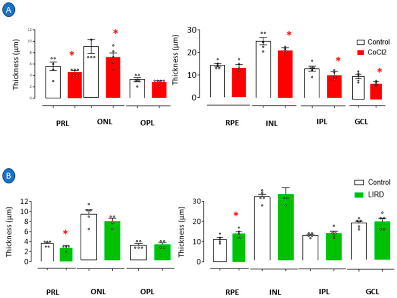
IMAGE

Figure 2

ID
ZDB-IMAGE-250701-39
Source
Figures for Rosa et al., 2025
Image
Figure Caption

Figure 2

Histologic analysis of retinopathy in zebrafish larvae. Retinopathy was induced in 3 dpf larvae through exposure to E2 0.5x medium at 28 °C with 0.5 mM cobalt chloride (CoCl2) for 72 h. Independent group of 3 dpf larvae was exposed to intense light at 8000 lux for 24 h (LIRD). Retinal layer thickness of CoCl2-exposed zebrafish larvae (A) and zebrafish larvae subjected to LIRD (B). * p < 0.05 compared to control group. Bars represent mean ± SEM. Dots represent individual data points. PRL: photoreceptor layer; ONL: outer nuclear layer; OPL: outer plexiform layer; RPE: retinal pigment epithelium; INL: inner nuclear layer; IPL: inner plexiform layer; GCL: ganglion cell layer.

Acknowledgments
This image is the copyrighted work of the attributed author or publisher, and ZFIN has permission only to display this image to its users. Additional permissions should be obtained from the applicable author or publisher of the image. Full text @ Pharmaceuticals (Basel)