IMAGE

Fig 2

ID
ZDB-IMAGE-250627-93
Source
Figures for Zamostiano et al., 2025
Image
Figure Caption

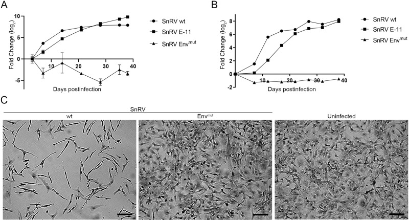
Fig 2 SnRV wt molecular clone is infectious.

(A, B) Kinetics of SnRV spread. BF-2 cells were infected with equal amounts - normalized by qRT-PCR with SnRV pol-derived primers (A) or RT activity (B) - of SnRV wt, SnRV Envmut, or SnRV from E-11 cultures (SnRV E-11). On the indicated days postinfection, the culture supernatants were sampled (140 µl) to quantify viral RNA levels by qRT-PCR (A), or harvested (10 ml) for pelleting the virions by ultracentrifugation to determine the pellets’ RT activity by SG-PERT (B). The fold change (log2) between each time point and (A) the initial time point (day 3 postinfection) or (B) the background sample (a pellet of a supernatant of naïve BF-2 culture) was calculated. Values represent the mean ± SD of three technical repeats. (C) Phenotypic changes in SnRV-infected BF-2 cells. Sub-confluent BF-2 cultures were infected (SnRV wt or SnRV Envmut), or not. The cultures were passaged for 18 days postinfection, stained with crystal violet, and imaged by light microscopy. Bar; 100 µm.

Acknowledgments
This image is the copyrighted work of the attributed author or publisher, and ZFIN has permission only to display this image to its users. Additional permissions should be obtained from the applicable author or publisher of the image. Full text @ PLoS Pathog.