IMAGE

Fig. 5

ID
ZDB-IMAGE-250618-7
Source
Figures for Li et al., 2025
Image
Figure Caption

Fig. 5

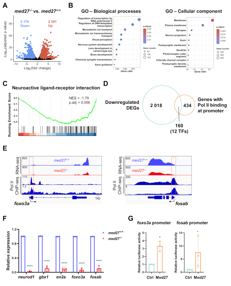
med27 LoF resulted in dysregulation of the transcriptional landscape related to neurodevelopment. A Volcano plot showing RNA-seq results comparing the transcriptomic profiles of med27+/+ and med27−/− larvae. RNA-seq was performed on pooled total RNA extracted from 7 dpf larvae from all three mutant lines (INS10, INS5, DEL5). 2 178 downregulated DEGs and 2 501 upregulated DEGs were identified (|Log2foldchange|>1, adjusted P < 0.05). B Top biological processes and cellular components identified by GO analysis of downregulated DEGs. The bubble size corresponds to the number of genes involved in each term, and the color intensity indicates the adjusted P value. C Gene set enrichment analysis (GSEA) plot showing enrichment of the neuroactive ligand-receptor interaction pathways among downregulated DEGs. D Venn diagram showing overlap of downregulated DEGs with positive Pol II binding at their promoter regions, based on Pol II ChIP-seq data (GEO Project ID PRJNA945049). TFs: transcription factors. E Bulk RNA-seq read counts of foxo3a (left) and fosab (right), along with the Pol II binding profiles at their promoter regions based on two Pol II ChIP-seq datasets (GEO Project ID PRJNA945049 and GSE175444). F RT-qPCR analysis of expression levels of identified overlapped TFs with well-established roles in brain and nervous system development. G Dual-luciferase reporter assays illustrating the activating effect of Med27 on promoter activities of foxo3a (left) and fosab (right). The assays were performed in zebrafish embryos. Ctrl: embryos injected with the pCS2 backbone vector; Med27: embryos injected with the pCS2-med27 expression vector. Error bars represent mean ± SD. Statistical analysis was performed using unpaired t-test (Fig. 5F) or Mann Whitney test (Fig. 5G). *: P < 0.05; ****: P<0.0001

Acknowledgments
This image is the copyrighted work of the attributed author or publisher, and ZFIN has permission only to display this image to its users. Additional permissions should be obtained from the applicable author or publisher of the image. Full text @ Cell Biosci.