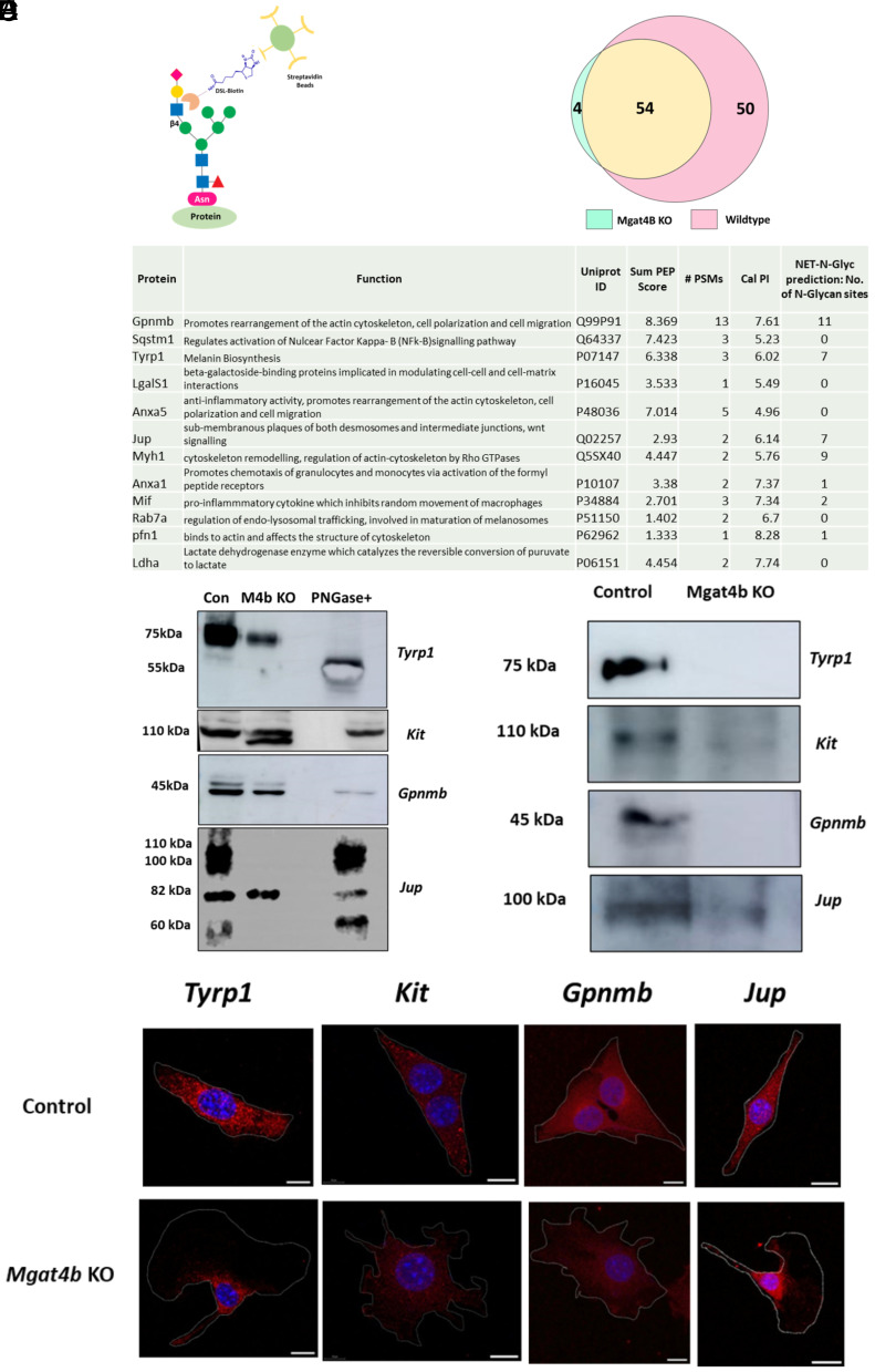
Differential proteomics reveal melanocyte specific target proteins of MGAT4B. (A, B) Schematic overview of the experimental strategy used to identify downstream targets of MGAT4B in melanocytes, Venn diagram depicting number of proteins identified by mass spectrometry analysis of the DSL-enriched fractions of Wildtype and Mgat4b KO proteins. (C) List of differentially enriched proteins in wildtype lysates with respect to Mgat4b KO along with their Uniprot ID, Sum PEP Score, PSMs, calc. PI and their functional profiling and predicted N-glycosylation sites by NetNGlyc—1.0 database. (D) Western blot analysis of Tyrp1, Kit, Gpnmb, and Jup in wildtype and Mgat4b knockout (M4b KO) lysate along with PNGase treated wildtype lysate. (E) Western blot analysis of Tyrp1, c-Kit, Gpnmb, and Jup following biotinylated DSL pull-down in wildtype and Mgat4b knockout (M4b KO) lysates. (F) Confocal microscopy images of immunocytochemistry using Tyrp1, Kit, Gpnmb, and Jup (red) on permeabilized wildtype and Mgat4b KO cells, counterstained by DAPI (blue). The overlay of both channels is depicted. [Scale bar, 50 µm (DSL- Datura stramonium Lectin].
Acknowledgments
This image is the copyrighted work of the attributed author or publisher, and
ZFIN has permission only to display this image to its users.
Additional permissions should be obtained from the applicable author or publisher of the image.
Full text @ Proc. Natl. Acad. Sci. USA
Your Input Welcome
Thank you for submitting comments. Your input has been emailed to ZFIN curators who may contact you if
additional information is required.
Oops. Something went wrong. Please try again later.