IMAGE

Fig. 1.

ID
ZDB-IMAGE-250611-68
Source
Figures for Sharma et al., 2025
Image
Figure Caption

Fig. 1.

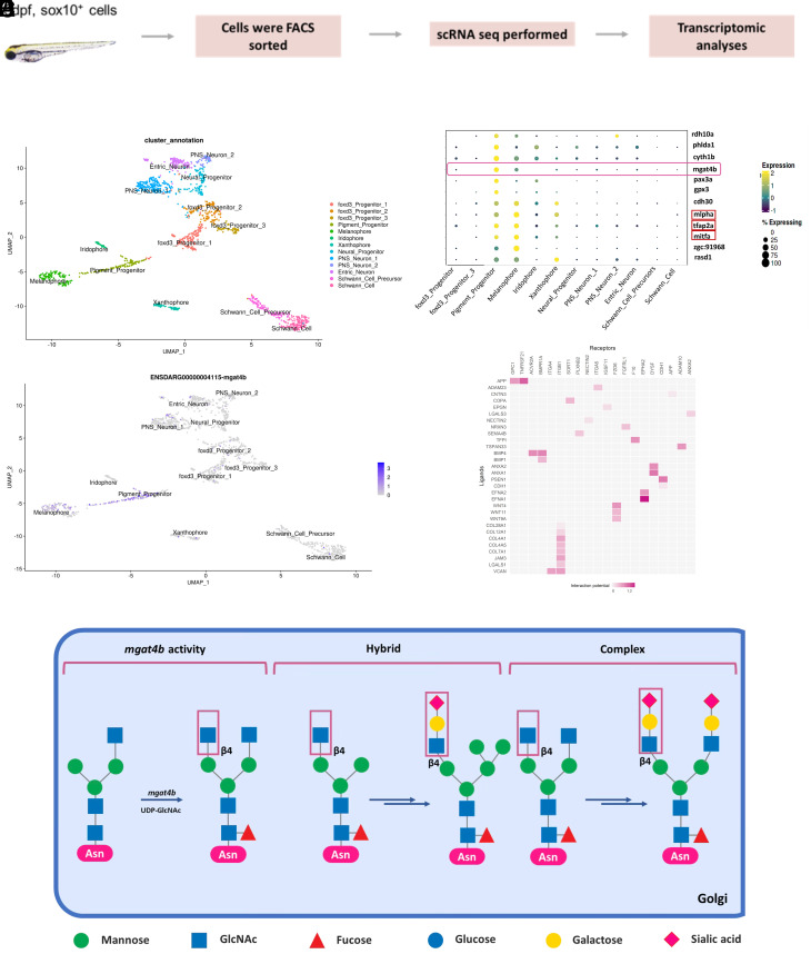
mgat4b is enriched in pigment progenitors and melanophores. (A) UMAP visualization of the Zebrafish Sox10+ve cells colored by the identified states. (B) Dot plot showing the top marker genes enriched in each cluster with the size showing the percent of cell expressing the gene and color showing the scaled mean expression value in each cluster (Wilcoxon–Mann–Whitney test with average log fold change > 0.25 and adjusted P value ≤ 0.05). List includes known cell-type marker genes (marked in red box) and new candidate markers. (C) UMAP plot shows enrichment of mgat4b in pigment progenitor and melanophore clusters. (D) Mammalian protein N-glycosylation starts in the endoplasmic reticulum and continues in the Golgi apparatus, where three mature types of N-glycan structures—high-mannose, hybrid, and complex—are formed. The enzyme MGAT4B transfers N-acetylglucosamine in a β1 → 4 linkage to α1 → 3-linked mannose, which initiates the creation of a specific N-glycan branch. This branch often includes a sialylated N-acetyllactosamine sequence, highlighting its distinctive features in glycan structure. (E) Heatmap of NicheNetR-identified ligand/receptor pairs indicating interaction potential between mgat4b+ve receiver cells and other sender cells sampled by scRNAseq of zebrafish Sox10+ve cells.

Acknowledgments
This image is the copyrighted work of the attributed author or publisher, and ZFIN has permission only to display this image to its users. Additional permissions should be obtained from the applicable author or publisher of the image. Full text @ Proc. Natl. Acad. Sci. USA