
|
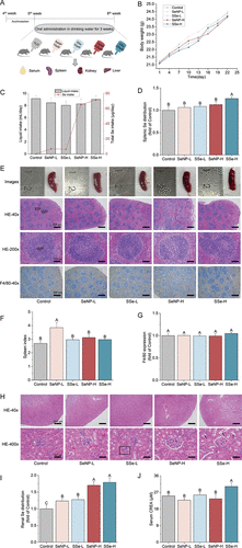
Fig. 3 Growth, splenic, and renal conditions of mice were altered by different Se-chemicals treatment. (A) Murine assay schematic diagram. (B) Body weight. (C) Liquid intake and Se intake for one mouse. L: low-Se group received drinking water containing 10 μM Se; H: high-Se group received drinking water containing 100 μM Se. (D) Splenic Se distribution. (E) Images of spleen (scale bar: 1 cm), HE staining (scale bar: 500 μm for 40×, 100 μm for 200×), and IHC of F4/80 (scale bar: 500 μm). (F) Spleen index. (G) Quantitative analysis of F4/80 expression in (E). (H) Images of renal HE staining. The rectangular box indicating denatured and shrunken glomeruli. The arrows pointing to deformed renal tubular epithelial cells. Scale bar: 500 μm for 40×, 50 μm for 400×. (I) Renal Se distribution. (J) Serum creatinine (CREA). The data represent the mean value ± SD. Different capital letters indicate significant differences among groups (p < 0.05).