IMAGE

Fig. 7

ID
ZDB-IMAGE-250609-54
Source
Figures for Chen et al., 2025
Image
Figure Caption

Fig. 7

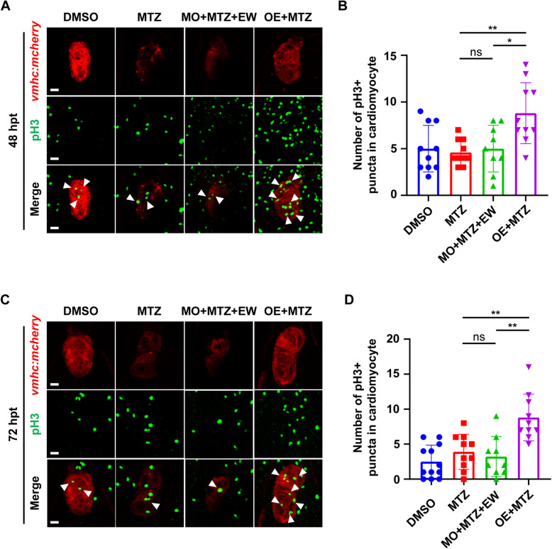
mvda affects the proliferation of cardiomyocytes. Immunofluorescent analysis of cardiomyocytes proliferation at 48 (A), 72 hpt (C) in mvda overexpression and knocking down group. White arrows indicate phosphorylated histone H3 positive (pH3 +) cardiomyocytes (CMs). Statistical analysis of cardiomyocytes proliferation at 48 (B), 72 hpt (D),. *, P < 0.05; **, P < 0.01. Scale bar, 20 μm

Acknowledgments
This image is the copyrighted work of the attributed author or publisher, and ZFIN has permission only to display this image to its users. Additional permissions should be obtained from the applicable author or publisher of the image. Full text @ Cell Regen (Lond)