Figure Caption
Figure 6
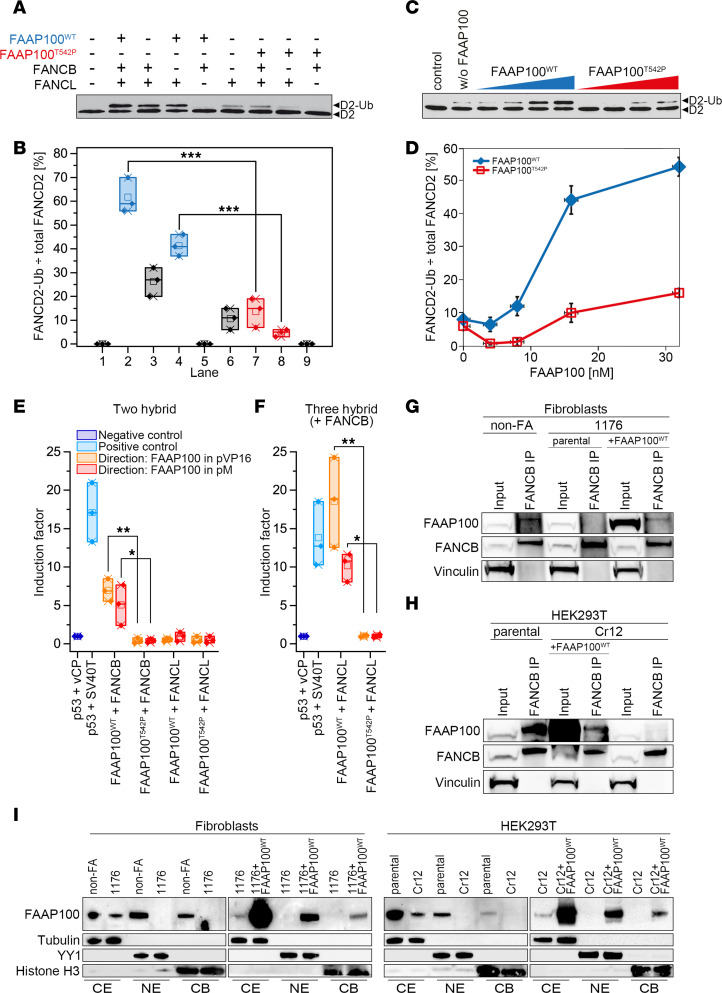
Ligase activity, interaction, and subcellular localization studies with FAAP100T542P.
(A) Reconstitution of FANCD2 monoubiquitylation using purified proteins, including HA-ubiquitin, UBE1, UBE2T, and FANCD2-FANCI complex and, as indicated, FAAP100WT or FAAP100T542P, FANCB, and/or FANCL. FANCD2 immunoblot: ◄D2-Ub, monoubiquitylated; ◄D2, nonubiquitylated. (B) Quantitation of results from A. FANCD2-Ub divided by total FANCD2 (percentage) indicates the ubiquitylation efficiency. Lane numbers are identical to those in A. Box plots: single value (♦), median (─), mean (□), IQR (─), minimum (x), and maximum (x) from 3 independent experiments. ***P < 0.001, by 1-way, repeated-measures ANOVA with post hoc Tukey’s test. (C) Titration of FAAP100WT (blue) and FAAP100T542P (red). For reaction mixtures, see A, including FAAP100WT or FAAP100T542P, FANCB, and FANCL. (D) Quantitation of results from C (see B and C for details). Data indicate the mean ± SD of 3 independent experiments. (E) In mammalian 2-hybrid assays, FAAP100WT, but not FAAP100T542P, interacted with FANCB. FAAP100 fused to the activation domain (orange) or the DNA-binding domain (red). Neither direction of FAAP100 fusion directly interacted with FANCL. Box plots: single value (♦), median (─), mean (□), IQR (─), minimum (x), and maximum (x) of 3 independent experiments. Induction factor, multiples of negative control. *P < 0.05 and **P < 0.01, by 1-way, repeated-measures ANOVA with post hoc Tukey’s test. (F) In mammalian 3-hybrid assays, FAAP100WT, but not FAAP100T542P, interacts with FANCL in both fusion directions in the presence of stably overexpressed FANCB. Controls, calculations, and statistical tests are the same as in E. *P < 0.05 and **P < 0.01, by 1-way, repeated-measures ANOVA with post hoc Tukey’s test. (G and H) Co-IPs from cells transfected with FANCB vector and exposed to MMC (40 ng/mL,16 hours). FAAP100T542P in 1176 cells and FAAP100L543_S551del in HEK293T clone Cr12 did not interact with FANCB. Transduced FAAP100WT rescued the pull-down of FAAP100 by FANCB. Vinculin was used as a loading control. (I) On subcellular protein fractionation, FAAP100T542P in 1176 fibroblasts and FAAP100L543_S551del in the HEK293T clone Cr12 were not detected in nuclear extracts (NE) or on chromatin (CB). Transduced FAAP100WT rescuedFAAP100 relocalization. CE, cytoplasmic extracts. Tubulin, YY1, and histone H3 were used as loading controls. Cells were exposed to MMC (40 ng/mL, 16 hours).
Acknowledgments
This image is the copyrighted work of the attributed author or publisher, and
ZFIN has permission only to display this image to its users.
Additional permissions should be obtained from the applicable author or publisher of the image.
Full text @ Journal of Clin. Invest.