IMAGE

Fig. 4

ID
ZDB-IMAGE-250602-105
Source
Figures for Lorenzini et al., 2025
Image
Figure Caption

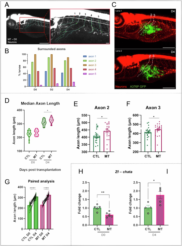
Fig. 4 Axonogenesis in zebrafish xenograft melanoma model.

A Representative images of a 3 dpf larva from NeuronStudio software. Five axons around the region of the SB were selected and manually traced in 3D using NeuronStudio to measure the axon length and to count the dendritic branching points. Scale bar = 250 μm/Scale bar = 50 μm. B The graph shows the percentage of larvae with axons 1–5 surrounding A375P cells. C Representative images of 7 dpf (D4) larvae with marked axon numbers 2 and 3. The images are taken using a confocal microscope. Scale bar = 250 μm. DG Axonogenesis was observed in the xenograft melanoma model. D The graph shows the average length of the five axons in every single larva in every condition on D0 and D4. n = 19–26. The measure of the axon length was performed using the NeuronStudio software. After manually tracing the axons and the dendritic branches, the software gave a summary with the length of every axon. E, F The graphs show the axon length on D4 of the axon number 2 (E) and axon number 3 (F), the most affected axons by the presence of A375P cells. Differences among groups were analyzed by two-tailed unpaired Student’s t test in (DF): *P-value < 0.05. n = 19–23. G The graph represents the paired analyses of the average of the five axons length of CTL and MT conditions on D0 and D4 as indicated. Differences among groups were analyzed by two-tailed paired Student’s t test in (G): *P-value < 0.05, **P-value < 0.01, ***P-value < 0.001, ****P-value < 0.0001. n = 19–22. The values in (DG) are expressed in μm. H, I Quantitative PCR analysis of the expression of the presynaptic cholinergic gene, choline acetyltransferase (chata), in the CTL and MT samples on D0 and D4 as indicated. Two-tailed unpaired Student’s t test was performed to evaluate the significance: *P-value < 0.05, **P-value < 0.01. n = 4–6. Results in (E, F) and (H, I) are expressed as means ± SEM. CTL = larvae injected with PBS. MT (melanoma transplant) = larvae transplanted with A375P GFP+ cells.

Acknowledgments
This image is the copyrighted work of the attributed author or publisher, and ZFIN has permission only to display this image to its users. Additional permissions should be obtained from the applicable author or publisher of the image. Full text @ Cell Death Discov