IMAGE

FIGURE 5

ID
ZDB-IMAGE-250530-108
Source
Figures for Balantzategi et al., 2025
Image
Figure Caption

FIGURE 5

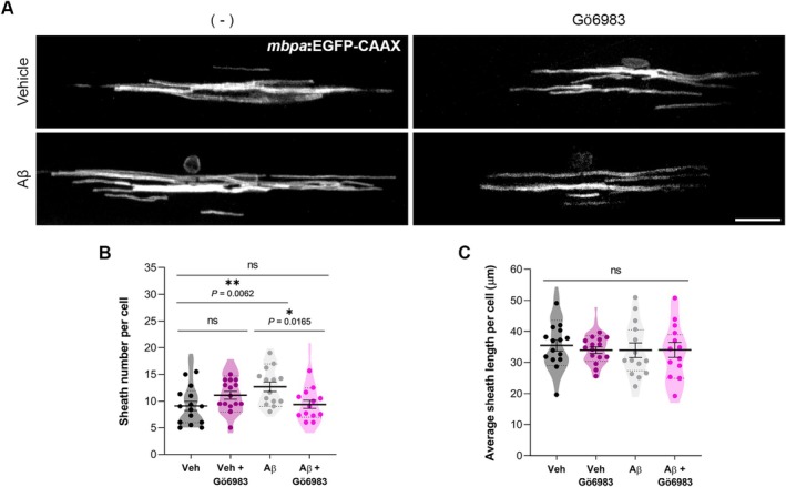
Aβ dysregulates dorsal myelin sheath number per oligodendrocyte via PKC activation. The mbpa:EGFP‐CAAX plasmid was injected into zebrafish embryos at the 1‐cell stage, followed by the administration of 10 μM of Aβ or its vehicle into the hindbrain ventricle of zebrafish larvae at 24 hpf. 500 nM Gö6983 treatments were performed at 48 hpf, and live imaging of myelin sheaths was performed at 5 dpf. (A) Representative fluorescent images of OLs in the spinal cord of live zebrafish larvae. (B) Histograms illustrating the sheath number per cell and (C) the average individual sheath length. Scale bar = 20 μm. Data are presented as means ± SEM; dots represent individual larvae and violin plots quantification of individual cells. *p < 0.05, **p < 0.01; Statistical significance was determined by two‐way ANOVA followed by Sidak's post hoc. n = 13–15 larvae per condition.

Acknowledgments
This image is the copyrighted work of the attributed author or publisher, and ZFIN has permission only to display this image to its users. Additional permissions should be obtained from the applicable author or publisher of the image. Full text @ Glia