
|
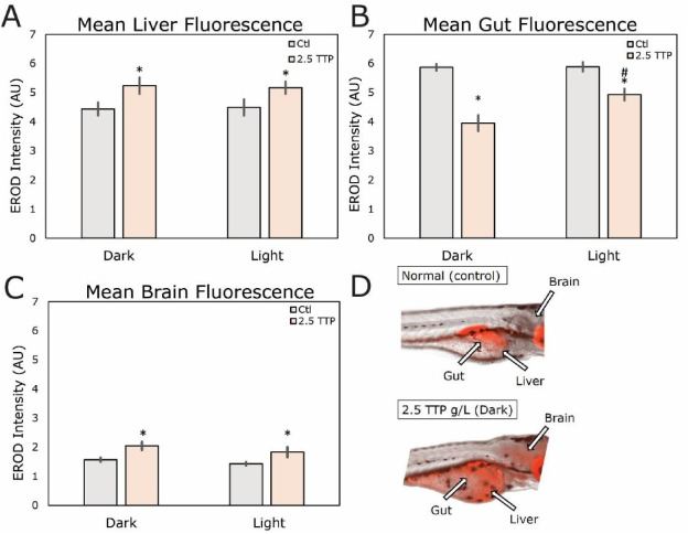
Fig. 2 At 3 dpf, embryos were exposed to 6-day leachate (dark or light) at 0 or 2.5 g/L in addition to six μL of 7-Ethoxyresorufin (7-ER). At 4dpf, embryos were evaluated for (D) EROD activity in the liver, gut, and brain. Activity was measured through the intensity of RFP fluorescence. The mean fluorescence intensity was measured and normalized to the background in order to get the (A) mean liver fluorescence, (B) mean gut fluorescence, and (C) mean brain fluorescence. T-tests were used to assess the significant difference between groups. n = 21–37 embryos per group. Asterisks (∗) indicate statistically significant changes from controls (p < 0.05). An octothorpe (#) indicates statistically significant changes from the dark group in the same concentration.