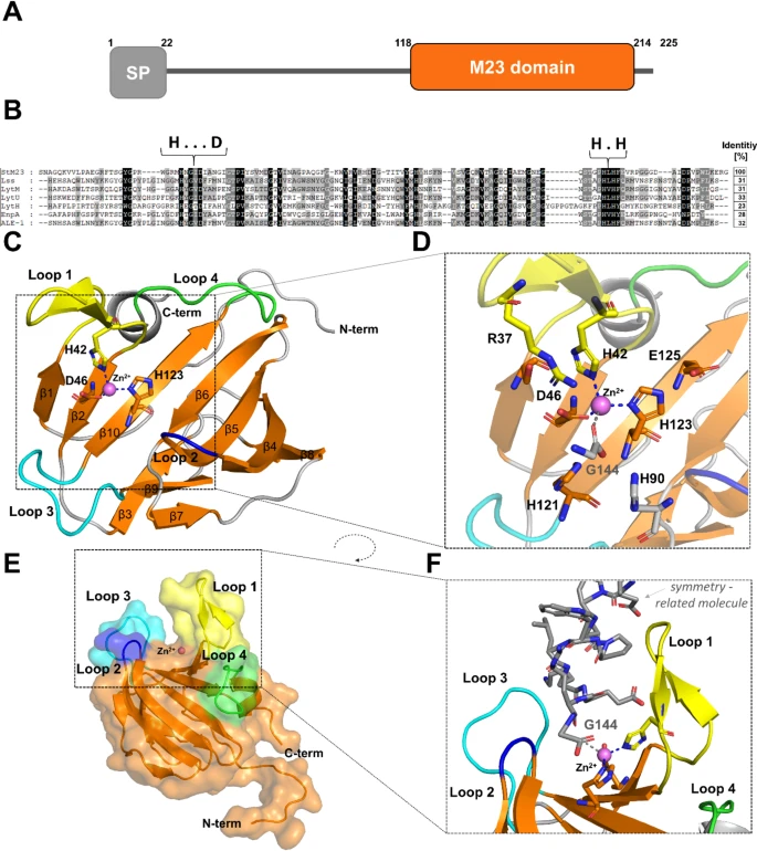
Fig. 1 Bioinformatic and structural analysis of StM23 protein (PDB ID:9GY1). (A) Domain composition of the native M23/37 peptidase identified in S. thermophilus NCTC 10353, highlighting key structural features.(B) Sequence conservation analysis of M23 peptidases, showing the sequence identity of StM23 compared to other related peptidases. Zinc-binding motifs HxxxD and HxH are marked. (C) The overall fold of the M23 peptidase with characteristic structure of two beta sheets (orange) and distinctive loops: loop 1 (yellow), loop 2 (blue), loop 3 (cyan), and loop 4 (green), with residues involved in zinc binding highlighted. Zinc ion is shown as a purple sphere. (D) A closer view of the active site architecture, emphasizing residues that play a crucial role in catalytic function and active site stabilization. (E) Surface representation of the StM23 peptidase structure, illustrating the characteristic groove and the zinc ion (Zn²⁺) position in the groove. (F) Close-up view of the groove with zinc ion coordination, highlighting the role of Gly144 (depicted in grey) in zinc coordination.
Image
Figure Caption
Acknowledgments
This image is the copyrighted work of the attributed author or publisher, and
ZFIN has permission only to display this image to its users.
Additional permissions should be obtained from the applicable author or publisher of the image.
Full text @ Sci. Rep.

