
|
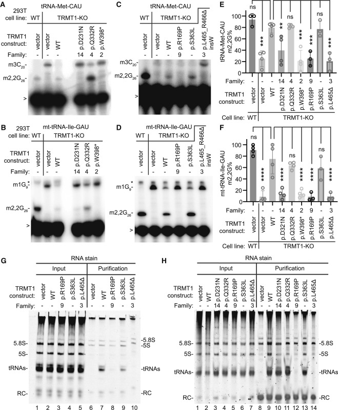
Fig. 5 TRMT1 protein variants exhibit defects in reconstitution of tRNA-modification activity and interaction with tRNAs (A–D) Representative primer extension gels to monitor the presence of m2,2G in tRNA-Met-CAU and mt-tRNA-Ile-GAU from 293T cell lines transfected with the indicated constructs. m3C20, 3-methylcytosine; m2,2G26, dimethylguanosine; m1G9, 1-methylguanosine. “>” points to oligonucleotide used for primer extension; asterisk denotes background signal. (E and F) Quantification of m2,2G formation by primer extension for the indicated tRNAs. Primer extensions were performed at least three times per variant, and error bars represent the standard error of the mean. Statistical analysis was performed using one-way ANOVA. ∗p ≤ 0.05, ∗∗p ≤ 0.01, ∗∗∗p ≤ 0.001, ∗∗∗∗p < 0.0001; ns, non-significant (p > 0.05). (G and H) Nucleic acid stain of RNAs extracted from the indicated input or purified TRMT1-FLAG samples after denaturing PAGE. The migration pattern of tRNAs, 5.8S, and 5S ribosomal RNA is denoted. The p.L465_R466Δ insW variant is denoted as L465Δ in (E)–(H).