IMAGE

Fig. 2

ID
ZDB-IMAGE-250514-41
Source
Figures for Mitra et al., 2025
Image
Figure Caption

Fig. 2

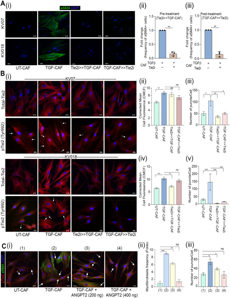
Tie2 plays essential role in induction as well as sustenance of TGFβ-induced myofibroblastic differentiation of CAF. (A) (i) Representative images and quantification of myofibroblasts frequency in UT-CAF and TGF-CAF. Tie2-inhibitor was added 1 h before TGFβ induction (Tie2i > > TGF-CAF) or 48 h after TGFβ induction (TGF-CAF > > Tie2i). Cells were quantified using ImageJ. (ii-iii) Frequency of αSMA stress fibre-positive cells were plotted for three different patient derived CAF. (B) (i) Representative images of Tie2 and pTie2 (Y992) in UT-CAF and TGF-CAF after Tie2-inhibition for 1 h before (Tie2i > > TGF-CAF) or 6 h after TGFβ induction (TGF-CAF > > Tie2i). (ii-v) Bar graph showing quantification of total Tie2 protein and pTie2 (Y992) puncta, calculated using ImageJ software. (C) (i) Representative images of αSMA and pTie2 (Y992) in UT-CAF, TGF-CAF or with increasing doses of ANGPT2 (200 ng/ml, 400 ng/ml) in the presence of TGFβ. Arrowhead indicates pTie2 (Y992) puncta. (ii) Bar graph showing cell frequency with αSMA stress fiber-positive CAF and (iii) pTie2 (Y992) expression by CAF in given conditions. Scale bar = 20 µm. *P < 0.05, **P < 0.01, ***P < 0.001

Acknowledgments
This image is the copyrighted work of the attributed author or publisher, and ZFIN has permission only to display this image to its users. Additional permissions should be obtained from the applicable author or publisher of the image. Full text @ J. Exp. Clin. Cancer Res.