
|
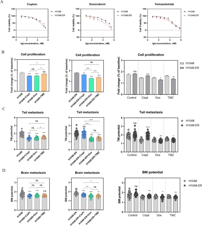
Fig. 6 Anti-proliferation and anti-metastasis effects of cisplatin, doxorubicin, and temozolomide on H1048 cells and H1048-ER cells in vitro and in vivo. A Growth curves of the parent cell line (H1048) and drug-resistant cell line (H1048-ER) under treatment with cisplatin (0.0096, 0.048, 0.24, 1.2, 6, 30, 150, and 300 μM), doxorubicin (0.0096, 0.048, 0.24, 1.2, 6, 30, 150, and 300 μM), or temozolomide (0.008, 0.04, 0.2, 1, 5, 25, 125, and 625 μM) for 24 h. Evaluation of B anti-proliferation, C anti-tail and D anti-brain metastasis activity of cisplatin (30 μM for proliferation assay, 0.6 pg/embryo for metastasis assay), doxorubicin (5 μM for proliferation assay, 1.09 pg/embryo for metastasis assay), and temozolomide (120 μM for proliferation assay, 3.88 pg/embryo for metastasis assay) against H1048 cells and H1048-ER cells in a zebrafish xenograft model at 3 dpt. ns no significance, *P < 0.033, **P < 0.002, and ***P < 0.001