
|
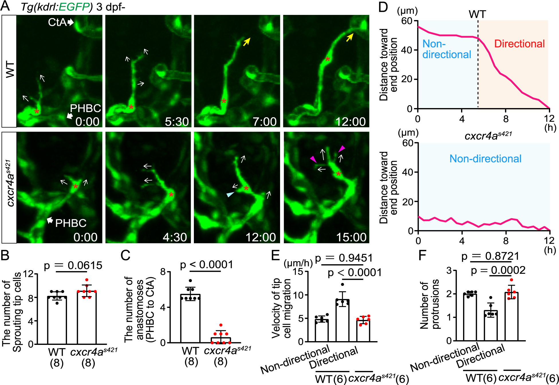
Fig. 2 Cxcr4a is important for directional migration to anastomotic targets (A) Time-sequential images of Tg(kdrl:EGFP) WT and cxcr4as421 sibling larvae (from 3 dpf). Asterisks indicate tip cell nuclei. In cxcr4as421 larvae, tip cells sprouting from the PHBC repeatedly extend (magenta arrowheads) and retract (blue arrowhead) their protrusions in various directions (arrows). They do not migrate toward the target vessel. (B) Graph shows the number of tip cells sprouting from the PHBC in WT and cxcr4as421 sibling larvae (from 73 hpf to 121 hpf). Data are mean ± s.d. (WT, n = 8 larvae; cxcr4as421, n = 8 larvae). Each dot represents an individual larva in this graph. (C) Graph shows the number of connections of tip cells sprouting from the PHBC to the CtAs or CCtAs in WT and cxcr4as421 sibling larvae (from 73 hpf to 121 hpf). Data are mean ± s.d. (WT, n = 8 larvae; cxcr4as421, n = 8 larvae). (D) Quantitative analysis of the data shown in (A). Minimum distance between the position of the tip cell nucleus at each time point and the position of the nucleus at 12 h in WT and cxcr4as421 sibling larvae. Connection to the target CtA occurs around 12 h in this WT larva as shown in (A). (E) Graph shows the time-averaged velocity of the tip cell nucleus in WT (non-directional and directional phases) and cxcr4as421 sibling larvae. Data are mean ± s.d. (WT, n = 6 cells in 4 larvae; cxcr4as421, n = 6 cells in 4 larvae). (F) Graph shows the time-averaged number of protrusions per tip cell in WT (non-directional and directional phases) and cxcr4as421 sibling larvae. Data are mean ± s.d. (WT, n = 6 cells in 4 larvae; cxcr4as421, n = 6 cells in 4 larvae). Scale bar: 10 μm. P values were determined by two-tailed Studentʼs t-test (B, C) and one-way ANOVA with Tukey’s test (E, F).