
|
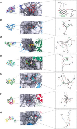
Fig. 7 Binding modes of TFLS compounds with target proteins. For target proteins lacking their native ligands, potential binding pockets were scored by SeeSAR during docking based on DoGSiteScorer. The yellow area (pocket 1) indicates the highest score, the red area (pocket 2) indicates the second highest, and the green area (pocket 3) indicates the lowest score. (A) Binding mode of epicatechin-(7,8-bc)-4β-(4-hydroxyphenyl)-dihydro-2(3H)-pyranone with PI3K. (B) Binding mode of quercetin with AKT1. (C) Binding mode of litchitannin A2 with ERK1. (D) The binding mode of dihydrochalcone-4′-O-β-D-glucopyranoside and ERK2; (E) The binding mode of procyanidin A2 and p38; (F) The binding mode of tamarixetin 3-O-rutinoside and mTOR. (G) The binding mode of litchiol B and JNK3.