
|
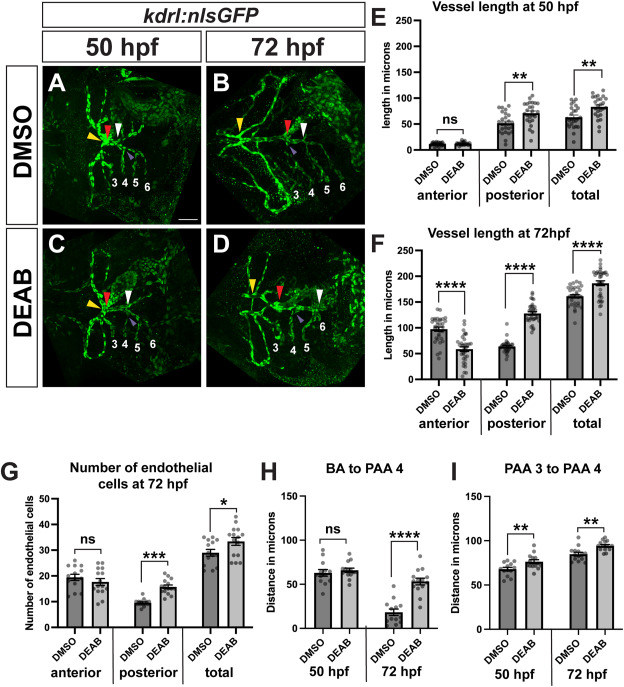
Fig. 9 The posterior ventral aorta domain is expanded in late RA-deficient embryos. (A–D) Representative images of immunostaining to detect kdrl:nlsGFP expression in endothelial cells at 50 hpf (A, C) and 72 hpf (B, D) in control and DEAB-treated embryos. Red arrowheads indicate center of the BA, which represents the boundary between anterior and posterior segments, yellow arrowheads indicate anterior end of fused ventral aorta, white arrowheads indicate posterior end of ventral aorta. Numbers indicate PAA positions. Purple arrowheads indicate the junction of PAA 4 and the ventral aorta. For all analyses, the distance between the yellow arrowhead and the red arrowhead represents the length of the anterior segment, and the distance between the white arrowhead and the red arrowhead represents the length of the posterior segment. (E) Quantification of ventral aorta lengths at 50 hpf in control (n = 25) and DEAB-treated (n = 25) embryos. (F) Quantification of ventral aorta lengths at 72 hpf in control (n = 33) and DEAB-treated (n = 34) embryos. (G) Quantification of distance between the OFT and PAA 4 at 50 hpf in control (n = 13) and DEAB-treated (n = 14) embryos and 72 hpf in control (n = 14) and DEAB-treated (n = 14) embryos. (H) Quantification of distance between PAA 3 and PAA 4 at 50 hpf in control (n = 13) and DEAB-treated (n = 14) embryos and 72 hpf in control (n = 14) and DEAB-treated (n = 14) embryos. (I) Endothelial cell number in ventral aorta segments at 72 hpf in control (n = 14) and DEAB-treated (n = 15) embryos. Scale bar is 50 μM. Error bars represent SEM. ∗∗∗∗ indicates p < 0.0001, ∗∗∗ indicates p < 0.001, ∗∗ indicates p < 0.01, ∗ indicates p < 0.05, ns = not significant. (For interpretation of the references to colour in this figure legend, the reader is referred to the Web version of this article.)
Reprinted from Developmental Biology, , Griffin, A.H.C., Small, A.M., Johnson, R.D., Medina, A.M., Kollar, K.T., Nazir, R.A., McGuire, A.M., Schumacher, J.A., Retinoic acid promotes second heart field addition and regulates ventral aorta patterning in zebrafish, , Copyright (2025) with permission from Elsevier. Full text @ Dev. Biol.