
|
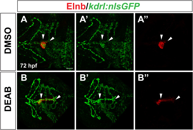
Fig. 8 The expanded posterior smooth muscle domain in late RA-deficient embryos surrounds an elongated posterior branch of the ventral aorta. Representative images of kdrl:nlsGFP expression detected by immunostaining in endothelial cells (A′, B′), Elnb expression in OFT smooth muscle (A″, B″), and the merged images (A, B). n = 16 for DMSO control, n = 24 for DEAB. White arrowheads indicate the posterior region of the ventral aorta in A′ and B′, the posterior “tail” of Elnb expression in A″ and B″, and the overlap of expression where the posterior Elnb domain surrounds the posterior region of the ventral aorta in A and B. Maximum intensity projections of a Z-stack are shown for each image. Scale bar is 50 μM, anterior is to the left in all images.
Reprinted from Developmental Biology, , Griffin, A.H.C., Small, A.M., Johnson, R.D., Medina, A.M., Kollar, K.T., Nazir, R.A., McGuire, A.M., Schumacher, J.A., Retinoic acid promotes second heart field addition and regulates ventral aorta patterning in zebrafish, , Copyright (2025) with permission from Elsevier. Full text @ Dev. Biol.