
|
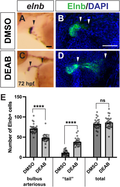
Fig. 7 OFT smooth muscle distribution is altered in late RA-deficient embryos. (A, B) ISH for elnb expression in the OFT in control (n = 12) and DEAB-treated (n = 19) embryos at 72 hpf. Black arrowheads indicate posterior elnb expression domain. (C, D) Representative images of immunostaining for Elnb protein in the OFT in control and DEAB-treated embryos at 72 hpf. Maximum intensity projections are shown. DAPI marks all nuclei in blue, Elnb expression is shown in green. White arrowheads indicate boundaries of the “tail”, or posterior Elnb expression domain. Maximum intensity projections of a Z-stack are shown for each image. (E) Quantification of Elnb expression in smooth muscle cells of the BA, “tail” region, and total cell number in DMSO (n = 26) and DEAB-treated (n = 27) embryos. Scale bars are 50 μM, anterior to the left in all images. Error bars represent SEM. ∗∗∗∗ indicates p < 0.0001, ns = not significant. (For interpretation of the references to colour in this figure legend, the reader is referred to the Web version of this article.)
Reprinted from Developmental Biology, , Griffin, A.H.C., Small, A.M., Johnson, R.D., Medina, A.M., Kollar, K.T., Nazir, R.A., McGuire, A.M., Schumacher, J.A., Retinoic acid promotes second heart field addition and regulates ventral aorta patterning in zebrafish, , Copyright (2025) with permission from Elsevier. Full text @ Dev. Biol.