
|
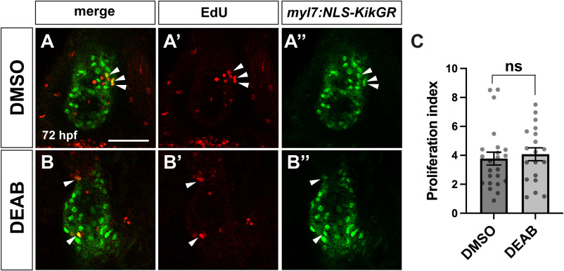
Fig. 6 Ventricular cardiomyocyte proliferation is unchanged in late RA-deficient embryos (A, B) Representative images of EdU staining in control DMSO (A) and DEAB-treated (B) embryos at 72 hpf. (A′, B′) EdU in red and (A″, B″) myl7:NLS-KikGR transgene expression in cardiomyocytes shown in green. Arrowheads indicate EdU+/NLS-KikGR + cardiomyocytes. Single confocal sections are shown. Scale bar is 50 μM. (C) Proliferation indices for DMSO (n = 23) and DEAB-treated (n = 19) embryos. (For interpretation of the references to colour in this figure legend, the reader is referred to the Web version of this article.)
Reprinted from Developmental Biology, , Griffin, A.H.C., Small, A.M., Johnson, R.D., Medina, A.M., Kollar, K.T., Nazir, R.A., McGuire, A.M., Schumacher, J.A., Retinoic acid promotes second heart field addition and regulates ventral aorta patterning in zebrafish, , Copyright (2025) with permission from Elsevier. Full text @ Dev. Biol.