
|
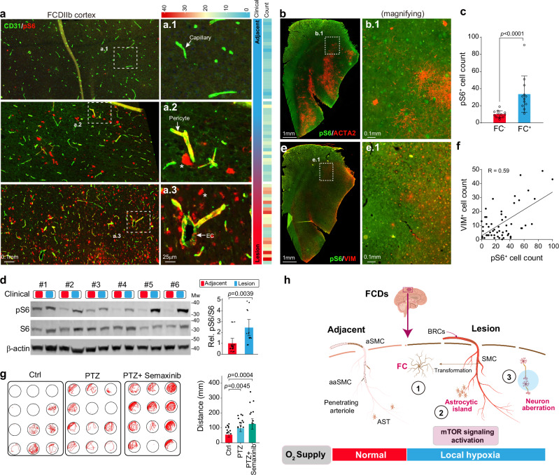
Fig. 5 Vascular malformation contributes to seizure pathogenesis. a Immunofluorescence (IF) staining for CD31 and pS6 for neocortex sections of FCDIIb patients. The right panel shows a local magnification of the sections within the dashed box. EC, endothelial cell. b, c IF staining for ACTA2 and pS6 (b) in lesion neocortex sections from FCDIIb patients and corresponding statistics (c). The cell number of 12 independent local sites (1 × 1 mm2) was counted. Data are presented as mean ± SD. P-value < 0.0001 by two-sided Mann-Whitney nonparametric test. Source data is provided as a Source Data file. FC, firework cell. d Western blot analysis of the indicated protein pS6 and S6 expression levels in the neocortex of FCDIIb patients. Both lesion and paired adjacent neocortex samples were used. pS6: phosphorylated S6 protein. n = 18. Data are presented as mean ± SD. P-value = 0.0039 by two-sided paired t-test. Mw, molecular weight. Source data is provided as a Source Data file. e, f IF staining for ACTA2 and VIM (e) in lesion neocortex sections from FCDIIb patients and associated statistics (f). The cell number of 54 independent local sites (1 × 1 mm2) was counted. P-value < 0.0001 of linear regression by F-test; R, Pearson correlation coefficient. Source data is provided as a Source Data file. g Behavioral testing of the improvement in PTZ-induced seizure activity in zebrafish treated with Semaxinib. PTZ pentylenetetrazole. n = 24. Data are presented as mean ± SD. P-value by two-sided Mann-Whitney nonparametric test. Source data is provided as a Source Data file. h Schematic model of how vascular malformation contributes to seizure pathogenesis. In the lesion region, these FCs fail to encircle endothelial cells as normal SMCs do, depleting the SMC pool and impairing the contractility of vessels (including arteries and arterioles SMC). This leads to the creation of a local hypoxia environment within the lesion neocortex, which, along with mTOR activation, further disrupts astrocyte and neuron functions. AST astrocyte, aSMC arteries SMC, aaSMC arterioles SMC.