
|
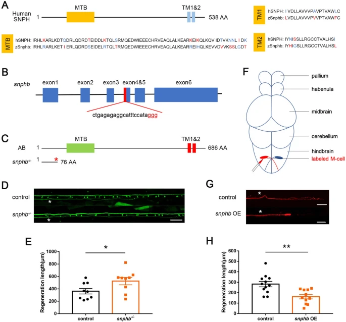
Fig. 1 snphb negatively regulates M-cell axon regeneration. A Zebrafish SNPHB is highly conserved with human SNPH in the MTB and TMs domains (alignment by http://multalin.toulouse.inra.fr/multalin/). B SgRNA target site located in the fourth exon of snphb. C Schematic of the protein sequence showed that snphb-/- zebrafish expressed SNPHB with deletion of MTB and TMs domains. D, E Representative images of control (T056) and snphb-/- M-cell axon regeneration results. Statistical results showed snphb knockout enhanced axon regeneration in zebrafish M-cells. White asterisk denotes the injury site. Scale bar, 50 μm, * P < 0.05, error bars indicate SEM. F Schematic of in vivo single-cell electroporation. G, H Representative images of overexpression backbone (control) and snphb OE axon regeneration results. Statistical results showed that snphb OE impeded axon regeneration in zebrafish M-cells. OE bg: 282.3 ± 26.12 μm; snphb OE: 161.5 ± 20.31 μm. White asterisk denotes the injury site. Scale bar, 50 μm, ** P < 0.01, error bars indicate SEM.