
|
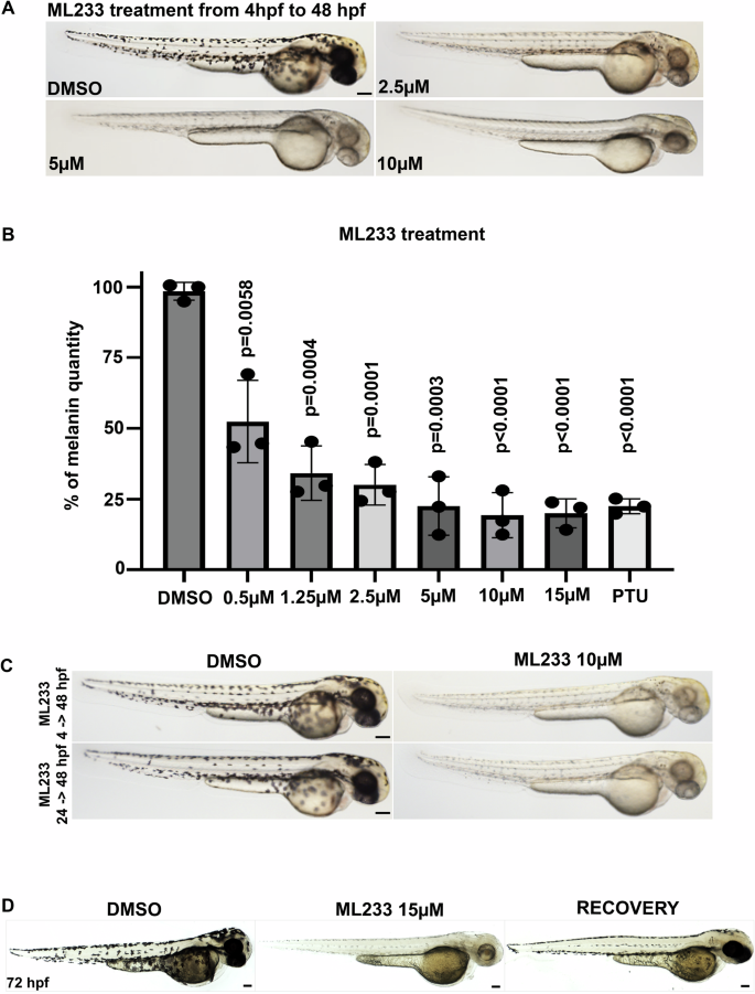
Fig. 2 A Skin pigmentation of DMSO- or ML233-treated embryos at 48 hpf after treatment between 4 and 48 hpf. B Melanin was quantified in control or ML233-treated embryos (n = 3 biological replicates with 3 technical replicates; ≥40 embryos/biological replicate). Significance is determined by t-test, two-tailed, unpaired. C Skin pigmentation of DMSO- or ML233-treated embryos at 48 hpf after treatment between 4 and 48 hpf (top) or 24 and 48 hpf (bottom). D Skin pigmentation of DMSO, ML233-treated embryos (treatment between 24 and 72 hpf) and ML233-treated embryos after recovery (treatment between 24 and 48 hpf) at 72 hpf. Scale bars: 100 μm. Error bars represent s.d.