
|
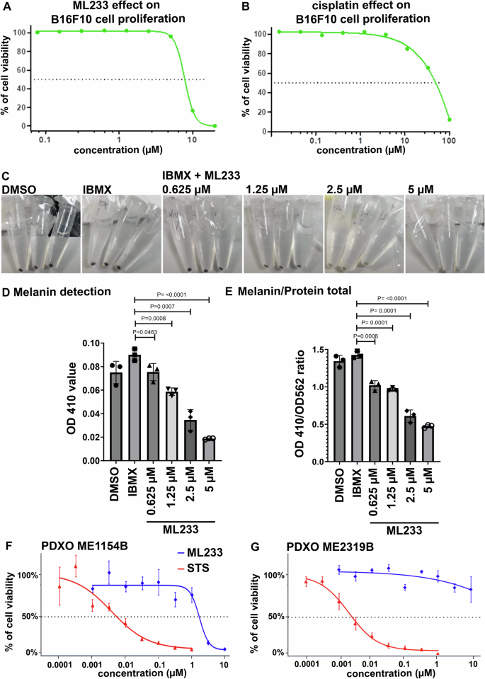
Fig. 6 Effect of ML233 (A) or cisplatin (B, positive control) treatment on B16F10 murine melanoma cell proliferation (n = 3). C Representative pictures of melanin expression in murine (B16F10) melanoma cells after treatment with DMSO, IBMX alone, or IBMX plus ML233. Melanin expression was quantified (D) and normalized (E) after treatment with DMSO, IBMX alone, or IBMX plus ML233 (n = 3). Significance is determined by t-test, two-tailed, unpaired. Cell proliferation in PDXO cellular models was quantified in ME1154B (F) or ME2319B (G) human melanoma lines after staurosporine (STS, positive control) or ML233 treatment (n = 3). Error bars represent s.d.