IMAGE

Fig. 8

ID
ZDB-IMAGE-250505-121
Source
Figures for Chen et al., 2025
Image
Figure Caption

Fig. 8

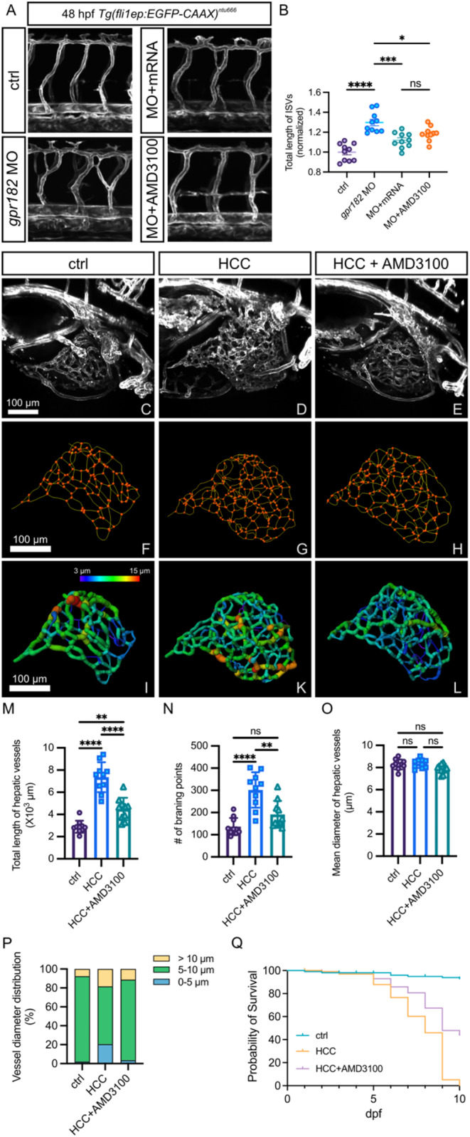
Inhibition of CXCR4 signaling normalizes the vasculature in gpr182 morphants and the liver of HCC model. (A) Confocal microscopy images of trunk vessels in 48-hpf Tg(fli1ep: EGFPCAAX)ntu666 control embryos, embryos injected with gpr182 MO, embryos injected with gpr182 MO and gpr182 mRNA, and embryos injected with gpr182 MO and AMD3100. (B) Quantitative analysis of total ISV length. (C-E) Confocal images of liver vasculature in control zebrafish, HCC model zebrafish, and AMD3100-treated HCC zebrafish at 7 dpf. (F-L) Three-dimensional reconstructions of liver vasculature in controls, HCC zebrafish, and AMD3100-treated HCC zebrafish. Vascular vessels and branching points are indicated by green and orange, respectively. (M-O) Quantifications of total hepatic vessel length, number of vascular branching points, and vessel diameters of liver vasculature. Data are presented as mean ± SD. One-way ANOVA analysis was applied. Not significant (ns); ****, p < 0.0001. (P) Vessel diameter distribution in controls, HCC zebrafish, and AMD3100-treated HCC zebrafish. (Q) Survival rates of controls, HCC zebrafish, and AMD3100-treated HCC zebrafish until 10 dpf

Acknowledgments
This image is the copyrighted work of the attributed author or publisher, and ZFIN has permission only to display this image to its users. Additional permissions should be obtained from the applicable author or publisher of the image. Full text @ Angiogenesis