
Fig. 2
- ID
- ZDB-IMAGE-250430-12
- Genes
- Publication
- Fragale et al., 2025 - Loss of Atoh8 Affects Neurocranial and Axial Skeleton Development in Zebrafish
- All Figures
- Figures for Fragale et al., 2025
|
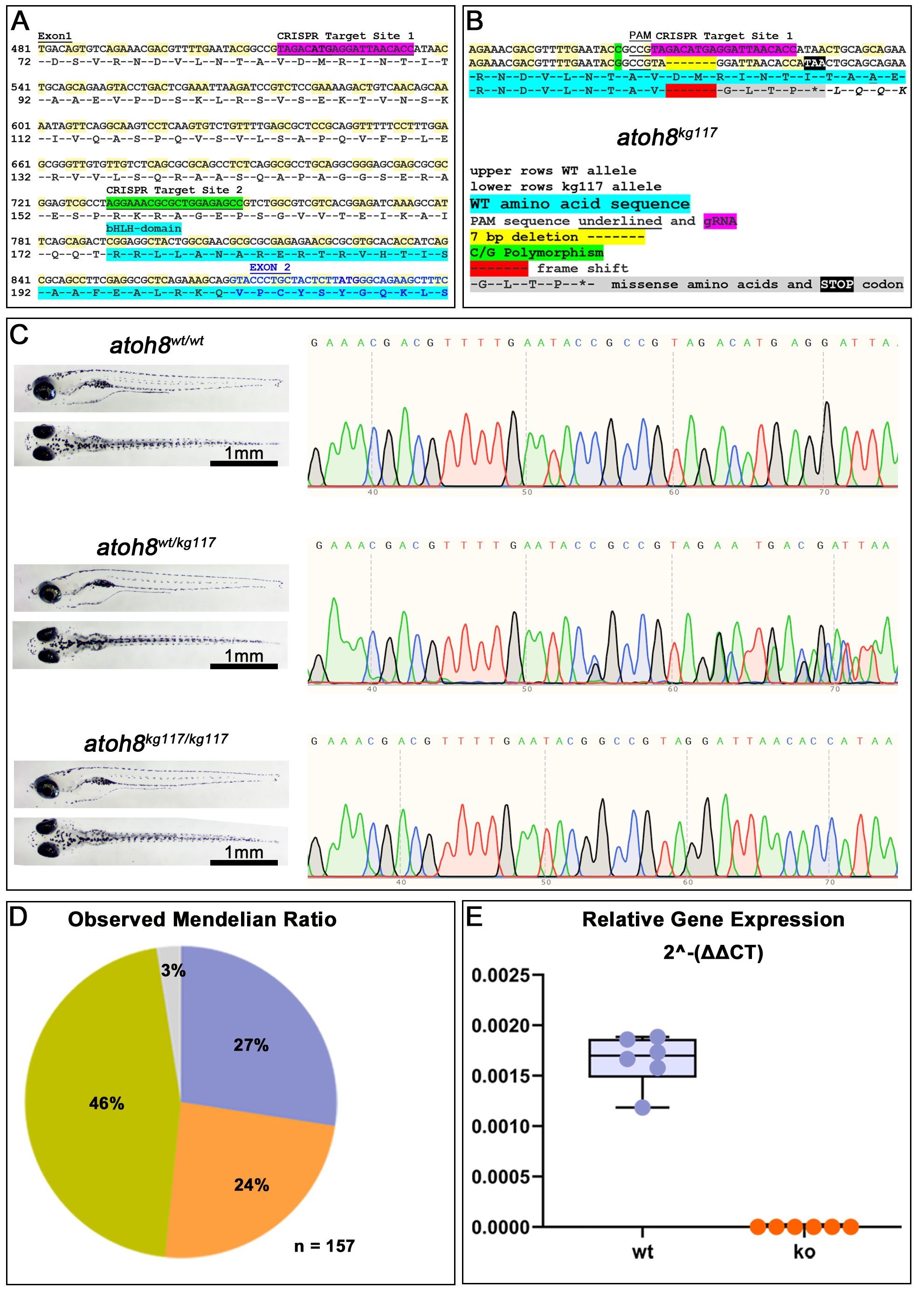
Fig. 2 CRISPR/Cas9-mediated mutagenesis. (A) Nucleotide and amino acid sequences showing the intended target sites for CRISPR/Cas9-mediated editing. Highlighted in light blue is the beginning of the functional bHLH domain, which is coded for partly in exon1 (black letters) and exon2 (blue letters) of the atoh8 gene. (B) Sequencing results in F1 progeny for the atoh8kg117 allele. The nucleotide deletion is highlighted in yellow in the DNA sequence and in red in the amino acid sequence of the protein. (C) Trace sequences for wildtype, heterozygous and homozygous siblings in the allele atoh8kg117. To the left: wildtype, heterozygous and homozygous sibling larvae shown in lateral (above) and dorsal (below) view. (D) The observed Mendelian Ratio is in very close correspondence with the expected Mendelian Ratio of 25% for wildtype (shown in lilac), 50% heterozygous mutants (green) and 25% for homozygous mutants (orange). The grey portion represents the percentage of larvae for which the genotype could not be determined. n = 157 sibling embryos of a atoh8wt/kg117 in-cross were used for genotyping and all subsequent analysis. (E) Relative gene expression in wildtype and homozygous mutant. Scale bars in (C): 1 mm. bHLH, basic helix-loop-helix; PAM, protospacer adjacent motif; gRNA, guide RNA; wt, wildtype; CRISPR/Cas9, clustered regularly interspaced short palindromic repeats/CRISPR-associated protein 9; ko, atoh8kg117/117.