IMAGE

Fig 2

ID
ZDB-IMAGE-250426-41
Source
Figures for Yin et al., 2025
Image
Figure Caption

Fig 2

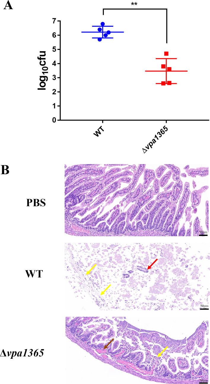
Analysis of the intestinal pathological section and bacterial colonization quantity in infant rabbits. (A) The amount of WT and Δvpa1365 colonized in the small intestine. **P ≤ 0.01. (B) Observations on the small intestine were challenged with PBS, WT, and Δvpa1365, respectively. The yellow arrow, red arrow, and brown arrow represented exfoliated intestinal villous epithelial cells, damaged lamina propria, and vascular congestion, respectively. Three infected tissues from each group were analyzed, and a representative image was presented for each case. Scale bars: 50 µm.

Acknowledgments
This image is the copyrighted work of the attributed author or publisher, and ZFIN has permission only to display this image to its users. Additional permissions should be obtained from the applicable author or publisher of the image. Full text @ Appl. Environ. Microbiol.