
|
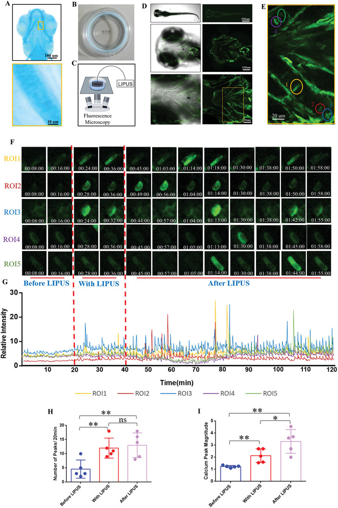
Figure 2
LIPUS promotes chondrocyte calcium oscillation of living zebrafish. A) Alcian blue staining to label the cranial cartilage structure of zebrafish juveniles. B) Zebrafish larvae were immobilized in a confocal dish with low‐melting‐point agarose. C) Schematic of the real‐time LIPUS‐processed calcium imaging under a confocal microscope. D) The whole fish, cranial and ceratohyal observed under a confocal microscope. E) 5 chondrocytes were circled with different colors as ROIs. F) The real‐time fluorescence intensity of the five ROIs. G) Calcium transient relative fluorescence intensity of ROIs. Quantification of the number H) (