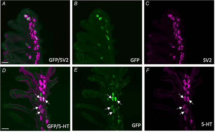
IMAGE

Figure 1

ID
ZDB-IMAGE-250423-67
Source
Figures for Reed et al., 2025
Image
Figure Caption

Figure 1 Characterization of GCaMP‐positive neuroepithelial cells (NECs) in the gill epithelium of transgenic elavl3:GCaMP6s zebrafish

Confocal imaging of immunohistochemical localization of GCaMP with NECs containing synaptic vesicle protein‐2 (SV2) and 5‐hydroxytryptamine (5‐HT). A, labelling with GFP (green) co‐localized with NECs containing SV2 (magenta). B and C, GCaMP and SV2 labelling shown separately. Scale bar = 20 µm in (A) to (C). D, labelling with GCaMP (green) co‐localized with NECs containing 5‐HT (magenta, arrows). E and F, GCaMP and 5‐HT labelling shown separately. Scale bar = 20 in (D) to (F). [Colour figure can be viewed at wileyonlinelibrary.com]

Acknowledgments
This image is the copyrighted work of the attributed author or publisher, and ZFIN has permission only to display this image to its users. Additional permissions should be obtained from the applicable author or publisher of the image. Full text @ J. Physiol.